Clinical and 3-Dimensional Outcomes in Novel Thread Lift Using Bidirectional Double Needles for Facial Skin Laxity and Double Chin Deformity
Won-Yong Lee1*
1 Re:ONE Clinic, 5th-floor SamA building, 435 Dosandaro, Gangnamgu, Seoul, Republic of Korea.
*Corresponding Author: Won-Yong Lee, Re:ONE Clinic, 5th-floor SamA building, 435 Dosandaro, Gangnamgu, Seoul, Republic of Korea, TEL: 82-2-540-0210 ; FAX: 82-2-930-0015; E-mail:leewonyong13@gmail.com
Citation: Won-Yong Lee (2019) Clinical and 3-Dimensional Outcomes in Novel Thread Lift Using Bidirectional Double Needles for Facial Skin Laxity and Double Chin Deformity. J Dermatol & Ther 3:113.
Copyright:© 2019 Won-Yong Lee, et al. This is an open-access article distributed under the terms of the Creative Commons Attribution License, which permits unrestricted use, distribution, and reproduction in any medium, provided the original author and source are credited
Received date: April 30, 2019; Accepted date: May 04, 2019; Published date: May 09, 2019.
Abstract
Background: Thread lifting with absorbable sutures has recently gained prominence for rejuvenating aging face. Considering laxity progresses in multiple directions, conventional one-dimensional thread with small barbs may have intrinsic limitations for lifting 3-dimensional (3D) facial contours harmoniously. A convergent bidirectional poly-L-lactic acid (PLA) suture was introduced which has straight needle at the ends and symmetric cones.
Objective: To evaluate the safety and efficacy of the bidirectional thread lifting regimens for counteracting facial skin laxity and double chin deformity with the application of 3D imaging system.
Methods: A 20-week, prospective, single-arm, clinical study was conducted for 38 Korean subjects with cutaneous aging signs. Based on the versatility of double needles, operators designed U-L shape combination patterns for facial laxity, and double straight and U-shape combination patterns for double chin. Objective assessments using a 5-point Global Aesthetic Improvement Scale (GAIS), 3D imaging system, and subjective satisfaction were recorded.
Results: At the 20-week follow up, 90% of subjects experienced GAIS score 4 (“much improved’) or better changes for facial skin laxity, and 72% showed score 4 or better changes for double chin tightening. Subjective assessments generally paralleled these patterns. The 3D analysis showed an upward migration of the volume of the lower face, which is clinically interpreted as facial lifting. No serious adverse effect was observed, and mild side effects were spontaneously resolved within a few days.
Conclusion: With this prospective study, these novel bidirectional thread lifting regimens were safe and effective for lifting and tightening ptotic soft tissues around the aging face.
Keywords:
rejuvenation, aging face, anti-aging, facial lifting, thread lifting.
Introduction
Biometric volume loss and ptosis of soft tissue are cardinal features of skin aging, manifested as flattened cheeks, noticeable nasolabial folds, sagging jaw line and double chin deformity (submental fat deposit) [1-3]. With recent trends toward less invasive techniques and minimal complication, internal suture suspension has been actively performed for tightening ptotic facial soft tissues [4,5]. A variety of barbed delayed absorbable threads are generally inserted with straight fashion to reposition sagging soft tissue in an upward direction. Considering ptosis and laxity progress in multiple directions, conventional one-dimensional thread lifting with small barbs may have intrinsic limitations for changing 3-dimensional (3D) facial contours harmoniously.
In this study, we applied a novel convergent bidirectional poly-L-lactic acid (PLA) suture with straight needles at both ends in the lifting procedure. Versatility of double needles allows an arbitrary angle to be made to strengthen and tighten specific areas compared with the straight only pattern [6]. For example, the jawline may become more definite with an angle pattern at the entry point. In addition, the symmetric cones on the suture provide a firm anchorage for the suture with both immediate and long-term efficacies. The PLA component has also been known to increase the volume and restore shapeliness of the face gradually [7,8].
One important issue in the field of cosmetic dermatology is the paucity of objective evaluation tools for post-treatment changes. We applied 3D imaging software technology to analyze post-treatment results in addition to surgeons’ assessments. In this study, we prospectively assessed the efficacy and safety of a novel thread lifting regimen for counteracting the descent of the soft tissues – facial skin laxity and double chin deformity.
Materials and Methods
Study design and subjects
A 20-week, prospective, single-arm study was conducted to investigate clinical outcomes of lifting using a novel convergent bidirectional thread with double needles for the aging face. Throughout the study procedures, the principles of the 1975 Declaration of Helsinki were followed, and informed consent was obtained from each participant. After receiving the treatment at the baseline, subjects visited the clinic at week 1 and 20. At every visit, photographs, image analysis, and participants’ subjective assessments were recorded. Thirty eight Korean women (aged 32-67 years, 18 Fitzpatrick III and 20 type IV) with mild to moderate aging signs of either facial skin laxity or double chin deformity were enrolled. Exclusion criteria included pregnancy, a history of keloidal scarring, and a history of cosmetic surgery or procedures for rejuvenation within 1 year prior to the study.
Threads
A convergent bidirectional absorbable suture (Silhouette SoftTM, Sinclaire Pharma, London, United Kingdom) was used in this study. It has straight needles at both ends and symmetric cones. It is made of PLA while its cones are made of lactide/glycolide (82%/18%). On each suture, there are two series of cones. Each has the same number of cones, which face in opposite directions towards the extremity of each suture, hence the term “bidirectional”. The cones are separated by tied knots, placed at 0.5 cm (8 cones) or 0.8 cm (12 cones), and 1.2 cm (16 cones) intervals from each other, free-floating between the knots. The central section of the suture is 2 cm long. This section is delimited by two knots and is free of cones.
Surgical techniques
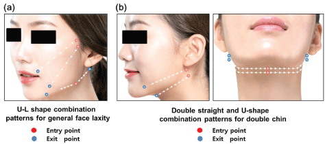
Before the procedure, local anesthesia was administered with 2% lidocaine with epinephrine (1:100,000) in each entry and exit point of all designs. Then, the operating field was painted with Betadine solution. Patients were laid down covering the chest with a sterile field. Then, operators designed vector patterns with a ruler. In this study, we used U-L shape combination patterns for facial laxity, and double straight and U-shape combination patterns for double chin (Figure 1).

Figure 1: Basic designs for the thread lifting. After patients were laid down covering the chest with a sterile field, the operators designed vector patterns with a ruler. In this study, we used (a) U-L shape combination patterns for general face laxity, and (b) double straight and U-shape combination patterns for double chin. Red dots: entry point; blue dots: exit point.
After creating the entry hole with an 18 G needle, operators performed the procedure with following sequences: 1) Insert the point of the first suture needle perpendicularly to the skin through the entry point to a depth of 5 mm into the subcutaneous tissue, then turn the needle horizontally and guide it through the subcutaneous tissue. 2) Extract the needle through the first exit point, pulling it gently. 3) When the last cone is inserted, stop the traction and start with the insertion of the second half of the suture. 4) Introduce the needle located at the second end of the suture, perpendicularly to the skin in the same entry hole. 5) Proceed in the direction of the second exit point and extract the needle with a gentle pull so that the cones are inserted in the tissue. After completing the entire suture, operators cut thread leaving the free ends long, and then pull the exposed ends of the sutures, so that the cones connect with the tissue to maintain the compression. Once adipose tissue compression has been completed, the free ends of the sutures at the exit points are trimmed. Oral cefaclor was prescribed for 3 days. Patients were not permitted to press directly or rub vigorously on the facial areas in which the threads were inserted during the first three months.
Outcome evaluation
After the treatment, follow-up visits took place at week 1 and 20. Digital photographic documentation using identical camera settings (EOS 600D®, Canon, Tokyo, Japan) and lighting conditions was obtained at each point. Two dermatologists not involved with procedures evaluated sequential photographs of the 38 patients in a randomized fashion to determine whether there was discernible clinical improvement. The efficacy was assessed by comparing photographs taken at each follow-up with those at baseline using a 5-point Global Aesthetic Improvement Scale (GAIS) (5: very much improved, 4: much improved, 3: improved, 2: no change, and 1: worse). Participants also subjectively reported their overall satisfaction degree using the following scale: “excellent,” “very good,” “good,” “fair,” or “poor.” Side effects were also recorded in detail. Pain sensation during treatments was reported on a 0-to-10 visual analogue scale (VAS) score (0: none, 10: extremely severe).
3D measurement
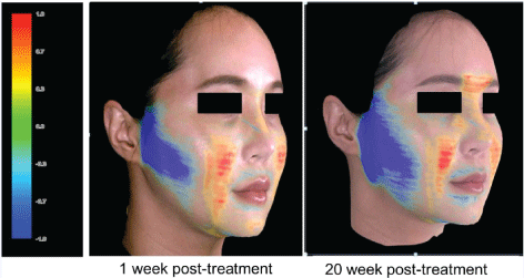
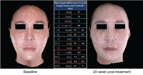
To provide quantitative data for facial skin laxity, 3D camera and software (Morpheus Co., Ltd., Seongnam, Republic of Korea) were also used. For six randomly selected patients, 3 images were taken from 3 different horizontal angles (the front, right, and left sides at an angle of 45Ëš) and then merged into a single 3D facial image. After anatomical landmarks were aligned, a series of lengths and angles were measured and compared. Quantitative volume measurements were also made using the 3D imaging software that compares the volume between before and after images. Superimposable 3D volumetric assessments allowed changes of volume to be reflected as color changes on the face.
Results
Objective assessment of efficacy
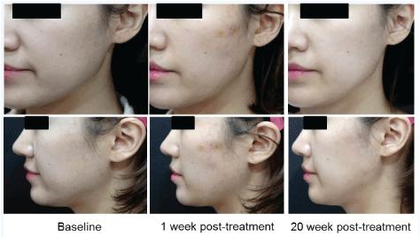
Clinical outcomes of thread lifting procedures are summarized in Table 1. Among 20 patients receiving lifting procedure for facial skin laxity, six subjects (30%) corresponded to GAIS score 5, and eleven subjects (55%) to score 4 after 1 week. At 20-week follow-up, eight (40%) received score 5, and ten (50%) score 4. Therefore, about 90% of subjects experienced “much improved” or better changes. Representative figures are shown in Figure 2. For 18 subjects receiving treatment for double chin tightening, 4 subjects (22%) received score 5, 7 subjects (39%) received 4, and 4 subjects (22%) received 3 at week 1. At week 20, 5 subjects (28%) received score 5, and 8 (44%) score 4. Therefore, 72% of subjects experienced “much improved” or better changes. Representative figures are shown in Figure 3.
Table 1: Clinical outcomes after thread lifting procedures. Surgeon’s objective assessment (Global Aesthetic Improvement Scale)
| General facial laxity (n=20) | Double Chin (n=18) | |||
|---|---|---|---|---|
| 1 week | 20 weeks | 1 week | 20 weeks | |
| 5 (Very much improved) | 6 (30%) | 8 (40%) | 4 (22%) | 5 (28%) |
| 4 (Much improved) | 11 (55%) | 10 (50%) | 7 (39%) | 8 (44%) |
| 3 (Improved) | 2 (10%) | 1 (5%) | 4 (22%) | 3 (17%) |
| 2 (No change) | 1 (5%) | 1 (5%) | 2 (11%) | 2 (11%) |
| 1 (Worse) | 0 (0%) | 0 (0%) | 1 (6%) | 0 (0%) |

Figure 2: Clinical photographs at baseline and 1 and 20 week post-operative follow-up points of a 48 year old female patient with general face laxity.

Figure 3: Clinical photographs at baseline and 1 and 20 week post-operative follow-up points of a 53 year old female patient with double chin deformity.
Participant’s subjective assessment
For both procedures, subjective assessments generally demonstrated comparable patterns. Detailed information is described in Table 2. At 20-week follow-up, seventeen patients (85%) reported “very good” or better changes for facial skin lifting. For double chin tightening, fourteen patients (78%) subjectively reported the comparable improvement.
Table 2: Subjective satisfaction after thread lifting procedures. Participant’s subjective assessment.
| General facial laxity (n=20) | Double Chin (n=18) | |||
|---|---|---|---|---|
| 1 week | 20 weeks | 1 week | 20 weeks | |
| Excellent | 5 (25%) | 6 (30%) | 5 (28%) | 7 (39%) |
| Very good | 8 (40%) | 11 (55%) | 7 (39%) | 7 (39%) |
| Good | 4 (20%) | 2 (10%) | 4 (22%) | 3 (17%) |
| Fair | 2 (10%) | 1 (5%) | 2 (11%) | 1 (6%) |
| Poor | 1 (5%) | 0 (0%) | 0 (0%) | 0 (0%) |
Image analysis based on 3D photographical sources and software program provided supportive data for facial skin laxity before and after thread lifting (Figure 4). Generally, the surface of central parts of the face became more prominent compared with that at baseline, while the surface of lateral sides of the face was depressed. To find possible quantitative anatomic markers for facial thread lifting, various angles and lengths were compared before and after treatments. Among them, curved lengths linking anatomical points in horizontal direction were increased, while those in vertical direction were decreased. These anthropometric quantitative indices may indicate the degree of tightening (Figure 5).


Safety profiles
For safety profiles, treatment was generally well tolerated, and it caused only minor discomforts. During treatment sessions, patients felt mild pain, with an average VAS score of 3.5. The most frequent complication was bruising, which occurred in 15 subjects (39%), followed by edema (13 subjects, 34%), mild asymmetry, and focal dimpling (6 patients, 16%) respectively. These side effects lasted for a maximum of 1 week and did not require further treatments. Significant adverse events such as nerve damage or foreign body granuloma were not observed in this study.
Discussion
In the field of aesthetic dermatology, absorbable suture suspension has attracted attention with the emergence of the so-called “lunch time” face lift [4,5]. To enhance the effect of the conventional one-dimensional thread insertion, we applied geometrically versatile, bidirectional sutures, which enable volume reposition and skin tightening in any area around face in the desired fashion. Furthermore, the repositioning of the facial soft tissues was measured objectively by 3D imaging system.
Results of our study demonstrated notable efficacy of these bidirectional thread lifting regimens. About 90% of patients demonstrated “much improved” or better improvements in the face lifting. These results suggest that the efficacy of this procedure may be better than that of other unidirectional thread lifting or non-invasive tightening devices [9-12]. Localized fat deposits in double chin deformity were also effectively redistributed. These lesions have been commonly a cause of discomfort and anguish, leading patients to undergo surgical procedures or mesotherapy to improve the cosmetic appearance with limited efficacies [2,13]. Combination patterns of the regimen may have contributed to the redefined distribution of the face by arbitrarily repositioning the redundant volume in desired directions. Especially, improvement with a firmer and a more regular contour in sagging jaw line with L and U designs were impressive for the elevation of both face and chin, respectively. Volume map changes by 3D images provided supporting data. No serious adverse effect was observed, and the pain was also not severe. Mild side effects reported were spontaneously resolved within a few days.
3D software has rarely been applied in the research of cosmetic procedures for soft tissue modulations. While craniofacial anthropometry and standardized photographs were previously used [14,15], they have intrinsic limitations in the analysis of soft tissue volume changes. Introduction of 3D technology as in this study has enhanced abilities to analyze post-treatment results in detail, allowing sophisticated analysis of volume repositions with clinical practicability [16-18]. More advanced technology dedicated to facial soft tissue analysis based on large clinical database would provide more benefits in clinical practice or related studies.
In addition to the versatility of bidirectional double needle, one of the distinguishing features of this suture is the existence of cones separated by tied knots. By having wide cross section areas with a 360º surface instead of conventional smaller barbs, they may provide excellent anchorage points for the suture in the subcutaneous tissue. While the cones slowly breaks down, capsules form around them. These capsules, which are not present with barbed sutures, histologically provide improved fixation points in the superficial adipose tissue [19,20]. Since the entire surface of the cones is smooth, no patient also experienced any prickling or pain sensation in this study.
PLA is a well-known polymer that has been used for many years as a biocompatible suture material in a variety of biomedical applications with FDA clearance [21]. It is generally classified as a safe material, belonging to the group of aliphatic polyesters. Ester bonds are hydrolyzed in the metabolic degradation process with the production of lactic acid, which is a substance normally present in the body. With its synthetic nature, it is generally well-tolerated, and no allergy testing is necessary [22,23]. In addition to instant lifting actions by tissue compression and skin traction with bidirectional cones, PLA components are known to stimulate the fibroblast and collagen production during the process of re-absorption [24]. This secondary regenerating action has a biostimulatory effect that leads to revolumization and sustained recontouring. Compared with other materials for sutures, PLA has a slower breakdown rate, with longer lasting benefits consequently [25].
There are some limitations in this study. First, all enrolled subjects are from same ethnic origins. Second, it is indicated for mild to moderate cutaneous laxities. For overabundant tissues, conventional surgeries such as lateral SMASectomy may be more suitable. Therefore, more standardized selection criteria based on 3D imaging tool should be adopted when establishing inclusion criteria. Third, a long-term follow-up for post-treatment changes would be required.
In conclusion, these bidirectional thread lifting regimens are safe and effective for lifting and tightening ptotic soft tissues around the face and chin, offering a non-surgical option for facial volume rejuvenation.
References
- Coleman SR, Grover R (2006) The anatomy of the aging face: volume loss and changes in 3-dimensional topography. Aesthetic surgery journal 26: S4-9.
- Co AC, Abad-Casintahan MF, Espinoza-Thaebtharm A (2007) Submental fat reduction by mesotherapy using phosphatidylcholine alone vs. phosphatidylcholine and organic silicium: a pilot study. Journal of cosmetic dermatology 6: 250-257.
- Helfrich YR, Sachs DL, Voorhees JJ (2008) Overview of skin aging and photoaging. Dermatol Nurs 20: 177-183. [crossref]
- Villa MT, White LE, Alam M, Yoo SS, Walton RL, et al. (2008) Barbed sutures: a review of the literature. Plastic and reconstructive surgery 121: 102e-108e.
- Garvey PB, Ricciardelli EJ, Gampper T (2009) Outcomes in threadlift for facial rejuvenation. Annals of plastic surgery 62: 482-485.
- Savoia A, Accardo C, Vannini F, Di Pasquale B, Baldi A, et al. (2014) Outcomes in thread lift for facial rejuvenation: a study performed with happy lift revitalizing. Dermatology and therapy 4: 103-114.
- Vleggaar D, Bauer U (2004) Facial enhancement and the European experience with Sculptra (poly-l-lactic acid). J Drugs Dermatol 3: 542-547. [crossref]
- Humble G, Mest D (2004) Soft tissue augmentation using sculptra. Facial plastic surgery : FPS 20: 157-163.
- Sulamanidze M, Sulamanidze G (2009) APTOS suture lifting methods: 10 years of experience. Clin Plast Surg 36: 281-306, viii. [crossref]
- Sulamanidze MA, Fournier PF, Paikidze TG, Sulamanidze GM (2002) Removal of facial soft tissue ptosis with special threads. Dermatologic surgery : official publication for American Society for Dermatologic Surgery [et al.] 28: 367-371.
- Alam M, White LE, Martin N, Witherspoon J, Yoo S, et al. (2010) Ultrasound tightening of facial and neck skin: a rater-blinded prospective cohort study. Journal of the American Academy of Dermatology 62: 262-269.
- Edwards AF, Massaki AB, Fabi S, Goldman M (2013) Clinical efficacy and safety evaluation of a monopolar radiofrequency device with a new vibration handpiece for the treatment of facial skin laxity: a 10-month experience with 64 patients. Dermatologic surgery : official publication for American Society for Dermatologic Surgery [et al.] 39: 104-110.
- Hexsel D, Serra M, Mazzuco R, Dal'Forno T, Zechmeister D (2003) Phosphatidylcholine in the treatment of localized fat. J Drugs Dermatol 2: 511-518. [crossref]
- Bartlett SP, Grossman R, Whitaker LA (1992). Age-related changes of the craniofacial skeleton: an anthropometric and histologic analysis. Plastic and reconstructive surgery 90: 592-600.
- Doual JM, Ferri J, Laude M (1997) The influence of senescence on craniofacial and cervical morphology in humans. Surgical and radiologic anatomy : SRA 19: 175-183.
- Aldridge K, Boyadjiev SA, Capone GT, DeLeon VB, Richtsmeier JT (2005) Precision and error of three-dimensional phenotypic measures acquired from 3dMD photogrammetric images. American journal of medical genetics. Part A 138A: 247-253.
- Weinberg SM, Naidoo S, Govier DP, Martin RA, Kane AA, et al. (2006) Anthropometric precision and accuracy of digital three-dimensional photogrammetry: comparing the Genex and 3dMD imaging systems with one another and with direct anthropometry. The Journal of craniofacial surgery 17: 477-483.
- Wong JY, Oh AK, Ohta E, Hunt AT, Rogers GF, et al. (2008) Validity and reliability of craniofacial anthropometric measurement of 3D digital photogrammetric images. The Cleft palate-craniofacial journal: official publication of the American Cleft Palate-Craniofacial Association 45: 232-239.
- de Benito J, Pizzamiglio R, Theodorou D, Arvas L (2011) Facial rejuvenation and improvement of malar projection using sutures with absorbable cones: surgical technique and case series. Aesthetic plastic surgery 35: 248-253.
- Huggins RJ, Freeman ME, Kerr JB, Mendelson BC (2007) Histologic and ultrastructural evaluation of sutures used for surgical fixation of the SMAS. Aesthetic plastic surgery 31: 719-724.
- Jabbar A, Arruda S, Sadick N (2017) Off Face Usage of Poly-L-Lactic Acid for Body Rejuvenation. J Drugs Dermatol 16: 489-494. [crossref]
- Tajbakhsh S, Hajiali F (2017) A comprehensive study on the fabrication and properties of biocomposites of poly(lactic acid)/ceramics for bone tissue engineering. Materials science & engineering. C, Materials for biological applications 70: 897-912.
- Hart DR, Fabi SG, White WM, Fitzgerald R, Goldman MP, et al. (2015) Current Concepts in the Use of PLLA: Clinical Synergy Noted with Combined Use of Microfocused Ultrasound and Poly-L-Lactic Acid on the Face, Neck, and Decolletage. Plastic and reconstructive surgery 136: 180S-187S.
- Goldberg D, Guana A, Volk A, Daro-Kaftan E (2013) Single-arm study for the characterization of human tissue response to injectable poly-L-lactic acid. Dermatologic surgery : official publication for American Society for Dermatologic Surgery [et al.] 39: 915-922.
- Rkein A, Ozog D, Waibel JS (2014) Treatment of atrophic scars with fractionated CO2 laser facilitating delivery of topically applied poly-L-lactic acid. Dermatologic surgery : official publication for American Society for Dermatologic Surgery [et al.] 40: 624-631.
