Imaging of Pedeatric Madiastinum
Kakarla Subbarao1*
1Department of radiology and imaging, KIMS, Secunderabad, India.
*Corresponding Author:Kakarla Subbarao, Department of radiology and imaging, KIMS, Secunderabad, India,; TEL::+91 40 6600 0066 ; FAX::+91 40 6600 0066;E-mail:subbaraokakarla25@gmail.com
Citation:Kakarla Subbarao(2016) Imaging of PedeatricMadiastinum. Cancer Prog Diagn1:104.
Copyright: :© 2016 Kakarla Subbarao, et al. This is an open-access article distributed under the terms of the Creative Commons Attribution License, which permits unrestricted use, distribution, and reproduction in any medium, provided the original author and source are credited.
Received date:October 06, 2016; Accepted date:October 31, 2016; Published date:November 9, 2016
Abstract
This is a review article on imaging findings in mediastinal pathology. Mediastinum is better studied on imaging methods such as plain radiography, MDCT, MRI and angiography.The characteristic findings of various mediastinal masses are described with a stress on plain radiography.Preoperative diagnosis can be made with imaging.
Key words
Pediatrics, mediastinal masses, plain radiography.
Introduction
Mediastinum is a median septum or partition containing mass of tissues and organs between the two pleural sacs.Anteriorly the sternum,posteriorly vertebral column, superiorly thoracic inlet and inferiorly diaphragm constitute its borders. Heart is considered to be in the anterior mediastinum. Aortic arch, descending aorta and pulmonary vessels are in the middle mediastinum.These structures can be identified in poster anterior and lateral chest radiographs and abnormal masses can be studied123. CT and MRI may help to study the matrix as well as the capsule, thus, giving a histological diagnosis4. FNAC / biopsyare conducted to confirm the diagnosis.The imaging methods are in the following tableTable I: Plain radiography of the chest – PA and lateral, MD CTMRI, Angiography, Pet CT
In children, ALARA principle, (as low as reasonably allowable) should be adopted so that minimal radiation is used as the children are very sensitive to radiation exposures.Mediastinum is divided into various compartments (Figure 1ab).Felson[1]and other authors have divided the mediastinum into various compartments and Felson’s method is followed in this study2.

Figure 1ab: a. Mediastinal compartments, b. A-Anterior, M- middle and P-posterior.
The following table lists the anterior mediastinal masses in children (Table II)
Table II: Enlarged thymus, Cystic Hygroma, Lymphoma, Teratodermoid
Thymomas are not generally encountered in children
In pediatric practice, most common mediastinal mass is the enlarged thymus.This may persist up to 4 years and rarely encountered even at 6-8 years[2]. Spontaneous regression is known and steroid therapy may help when the child is having respiratory problems (Figure 2ab).Radiologically, several signs have been described including the sail like density in the paratracheal area (Figure 3ab).

Figure 2ab :a- Persistent thymus, b-Post steroid therapy

Figure 3ab :a-Enlarged thymus, b-Sail like sign.
A wavy sign has been described where the costal cartilages produce an impression on the thymus (Figure 4ab-8abc).

Figure 4ab : a-Enlarged thymus – wavy sign, b-Compression by costal cartilages anteriorly
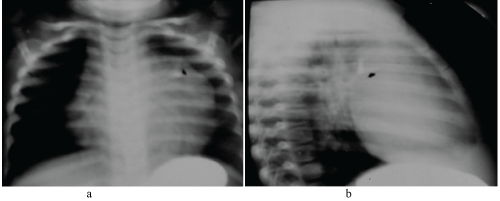
Figure 5ab :Teratodermoid - Note Calcification in the anterior mass

Figure 6ab :Large anterior mediastial mass due to lymphoma.

Figure 7ab: a-Lymphoma, b- 2 Months later with effusion.

Figure 8abc: ab- Right anterior mediastinal mass due to lymphoma, c- osteoarthropathy of the lower limbs.
The common anterior mediastinal masses, generally go by the eponem (4Ts)
-Thymus
-Thyroid
-Teratoma
-Terrible lymphoma
The masses in the middle mediastinum are listed in the table III:Lymph nodes, Bronchogenic cyst, Vascular Esophageal, Hernia
The most common middle mediastinal mass is due to enlarged lymph nodes
Theetiologies are listed in table IV: Tuberculosis, Sarcoid, Lymphoma, Leukemia, Infectiousmononucleosis, Pseudo lymphoma, Castleman’s disease, Angioimmuno lymphadenopathyFigure 9

CT imaging is performed to find out various sites of lymphadenopathy

Figure 10ab :Hodgkin’s lymphadenopathy, b-CT right paratracheal and para-aortic adenopathy

Figure 11abc :Lymphoma at various stages
Figure 10ab, Figure 11abcNext entity is sarcoidosis. Although,sarcoidosis is not very common in India and yet several cases have been reported in all ages[3]. Right paratracheal and bilateral hilar lymphadenopathy is characteristic. Unilateral hilar adenopathy may also be encountered[4].Figure 12

Figure 12 :16 yr old - Sarcoidosis with hilar lymphadenopathy .
Vascular rings and slings
These are often produce mediastinal widening simulating masses. Of these, right sided aorta with or without associated congenital heart disease is common. Although, plain films are classical as there is no normal aortic shadow on the left and an abnormal shadow is seen on right side of the trachea[5]. Normally, the trachea is deviated to the right by the presence of aortic knob. In right side aorta5, the trachea is deviated to the left (Figure 13a).

Figure 13a :Right sided aorta
Esophagogramconfirms the nature of the aorta. Angiogram is rarely necessary except when congenital heart disease is suspected (Figure 13bc).

Figure 13bc :Esophagogram with right sided aorta
Multidetector CT Angiogram is the latest investigation to be performed to study the cardiovascular structures (Figure 14ab)

Figure 14ab :a-Esophagogram, b-CT angiogram - Right sided aorta.
Next entity in the middle mediastinum comprises duplication cysts. Duplication cysts are congenital in nature and they may present as solid lesion on radiography (Figure 15ab).

Figure 15ab :Esophgeal duplication cyst.
If they communicate with the respiratory / esophagus tract6. They may contain fluid / air[6]. CT often helps in identifying the nature of contents (Figure. 15c)

Figure 15c :CT showing fluid in the cyst.
Observe alarp principle as far as possible to avoid radiation exposure in children[7]. CT of the chest is often avoided and replaced by ultrasonography or magnetic resonance imaging. The indications are mentioned in table V
Table V - Indications for CT Chest
Table V:Identify mediastinal mass, To differentiate between solid and cystic lesions, To identify calcification, To identify fat, To study cardiovascular system
Most of the posterior Mediastinal masses are neurogenic and include the following:
Table VI - Neurogenic Masses
Table VI:Neuroblastoma, Ganglioneuroma, Neurofibroma, Neurenteric cyst, Meningocele (Intrathoracic)Figure 16abc, Figure 17abc Neuroblastoma constitutes 75% of neurogenic tumors78. Next in order include ganlionic origin, ganglioneuroma, ganglioneuroblastoma[8]. Neurofibromas and schwannomas are uncommon in children. Figure 17 d The other masses include the following: Lymphoma, Extramedullary hemopoiesis mass, Hernia, Ectopic kidneyFigure 18

Figure 16a :6 yr M – Neuroblastoma.

Figure 16bc :CT neuroblastoma showing calcifications.

Figure 17abc :a-AP, b-Lateral, c-Esophagogram – Neuroblastoma with calcifications.

Figure 17d :CT necrotic centre in neuroblastoma.

Figure 18a :Oblong mass in ganglioneuroma.

Summary
Mediastinal Lesions in children are ideally studied by conventional radiographs. Where ever indicated advanced imaging methods are adopted. CT is minimally used to avoid excesive radiation. As far as possible ultrasonography and MRI studies should be replaced in the place of CT. Divisions of mediastinum help in identifying various masses.
References
- Felson B (1968) More chest roentgen signs and how to teach them. Annual Oration in memory of L. Henry Garland MD 1903-1966. Radiology 90: 429-441. [crossref]
- Felson B (1969) The mediastinum. SeminRoentgenol 4:41-58.
- Han BK, Babcock DS, Oestreich AE (1989) Normal thymus in infancy: sonographic characteristics. Radiology 170: 471-474. [crossref]
- Brown LR, Aughenbaugh GL (1991) Masses of the anterior mediastinum: CT and MR imaging. AJR Am J Roentgenol 157: 1171-1180. [crossref]
- Kuhlman JE, Fishman EK, Wang KP, Zerhouni EA, Siegelman SS, et al. (1988) Mediastinal cysts: diagnosis by CT and needle aspiration. AJR Am J Roentgenol 150: 75-78. [crossref]
- Predey TA, McDonald V, Demos TC, Moncada R (1989) CT of congenital anomalies of the aortic arch. SeminRoentgenol 24: 96-113. [crossref]
- Padovani B, Hofman P, Chanalet S, Taillan B, Jourdan J, et al. (1992) Intrapericardial bronchogenic cyst: CT and MR demonstration. Eur J Radiol 15: 4-6. [crossref]
- Sakai F, Sone S, Kiyono K, Maruyama A, Ueda H, et al. (1992) Intrathoracic neurogenic tumors: MR-pathologic correlation. AJR Am J Roentgenol 159: 279-283. [crossref]