Basic Histopathology of Breast Lesions
Dr. Somil Singhal 1*
1Post Graduate, Department of Pathology, S. Nijalingappa Medical College and HSK Hospital, Bagalkot, Karnataka, India.
*Corresponding Author:Dr. Somil Singhal, Post Graduate, Department of Pathology, S. Nijalingappa Medical College and HSK Hospital, Bagalkot, Karnataka, India. Tel: 083542 35360; Fax: 083542 35360; E-mail:somil15feb@gmail.com
Citation: Dr. Somil Singhal (2023)Basic Histopathology of Breast Lesions. Cancer Prog Diagn 7: 133.
Received: February 28, 2023; Accepted: March 29, 2023; Published: March 31, 2023.
Introduction
The clinical presentation of breast diseases can be multiple, including breast lump, breast “lumpiness,” nipple discharge, pain and redness of the overlying skin, or axillary lymph node enlargement. Nowadays, many breast lesions are asymptomatic, being detected by imaging or breast screening or as incidental findings during surgical removal for a different lesion. In general, the pre- sensation may give some hints to the nature of the underlying pathology. A solitary breast lump may represent either a benign or malignant breast tumor; typically, benign breast tumors are not fixed to the underlying structures, being freely mobile within the breast parenchyma, and palpa- tion or imaging shows rounded borders. A malignant tumor, on the contrary, shows irregular borders, and the tumor desmoplasia may render the mass firm too hard on palpation and appear fixed to the underlying parenchymal tissue or the overlying skin. Malignant breast masses are usually painless. Breast “lumpiness” gives a sensation of many small nodularity’s upon palpation, and this is characteristic of fibrocystic changes with the multiple small cysts being felt. There may be associated pain, which typically is related to the menstrual cycle. Nipple discharge is an uncommon and alarming symptom, particularly when the discharge is blood stained. Nipple discharge can be multiorificial or uniorificial. The former usually occurs when there is diffuse change in the breast, as occurs in fibrocystic changes, particularly when there is duct ectasia, when the secretions are accumulated within the ductal system, whereas in uniorificial discharge, there is usually an intraductal lesion or growth. The typical example in this situation is a papillary lesion. Both benign (duct papilloma) and malignant (papillary carcinoma) papillary lesions may give rise to blood-stained discharge. Pain and redness of the overlying skin are characteristic symptoms for acute inflammation or abscess of the breast. Rarely patients with breast cancer may present not with a clinicoradiological breast mass but with enlarged axillary lymphnodes; such cases are referred to as ‘occult pri- mary’ and warrant a diligent search for the primary tumor. Increasingly asymptomatic breast lesions are detected as a result of mammographic screen- ing, either because of architectural distortion or calcifications on imaging. The lesions may range from benign fibrocystic changes, radial sclerosing lesions and columnar cell changes to atypical lesions and tumor precursors, notably flat epithe- lial atypia (FEA) or atypical epithelial hyperpla- sia, to carcinoma in situ or small invasive cancers.
Inflammatory Breast Lesions
Mastitis or inflammation of the breast is uncommon. Mastitis can be divided into acute and chronic, and acute mastitis may be associated with abscess formation. Acute mastitis usually occurs at the postpartum period, when the lactating breast tissue is swollen, and sometimes the ducts are obstructed, with inspissation of the secretion.
Figure2.1:Breast tissue with chronic inflammatory cells within a congested background. There is also accumulation of inflammatory cells within the ductal lumen
.
In addition, breast-feeding may cause trauma and cracks to the nipple, resulting in ascending infec- tion of the commensals either originating from the skin or from the suckling baby’s oral cavity. More severe cases would result in abscess forma- tion. Histologically, there is infiltration of acute inflammatory cells, mostly neutrophils within the breast parenchyma. When there is abscess forma- tion, significant necrosis is present with collec- tion of necrotic debris and the exudates; at the same time, inflammatory reaction will result in granulation tissue formation and fibrotic tissue around the collection (Fig. 2.1). Duct ectasia is a common cause of chronic inflammation. In some cases of fibrocystic changes, the partially or totally blocked ducts may be distended by the accumulation of secretions from the lobules, resulting in cyst formation, and extravasation of the contents into the breast parenchyma will incite inflammatory response. This type of inflammation is aseptic and tends to be chronic, and sometimes, patients may present with nipple discharge. The histologic changes include infiltration of chronic inflammatory cells, including lymphocytes, plasma cells, and histiocytes; granulomas can sometimes be seen. Fat necrosis is a specific type of inflammatory change that occurs in the breast due to trauma or surgical procedure. Traumatic injury to the breast tissue causes disruption of the adipocytes, resulting in the release of the lipid into the stroma, eliciting an inflammatory response, with mostly histiocytes ingesting the fat. In addition, there is also fibrotic tissue reac- tion, resulting in the formation of a dense fibrotic scar that is hard on palpation, thus mimicking carcinoma on clinical examination. Depending on the stage of the evolution of the lesion, fat necrosis may present as a palpable nodule or just a focal area of pain. As a nodule, fat necrosis is often firm, and there may be skin retraction, thickening, or tethering. At ultrasound, in acute fat necrosis, there is an area of hyperechogenic- ity, which may have a central decrease in echo- genicity. For more long-standing lesions, fat necrosis forms a circumscribed to ill-defined mass and may show posterior acoustic shadow- ing. At mammography, especially when there is calcification or a spiculated mass, fat necrosis can be indistinguishable from carcinoma. Lipid cysts on mammography are diagnostic of fat necrosis. Similar effects could be seen in cases with breast augmentation, as some of the aug- mented materials may penetrate the breast paren- chyma, eliciting a similar inflammatory response.
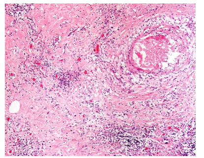
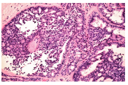
Figure 2.2: Granulomatous mastitis showing well-formed granulomas composing of epithelioid histiocytes and occasional Langhans giant cells. No caseation necrosis is seen. Alymphocytic infiltrate is noted in the background.
Chronic mastitis due to specific microorganisms is rare, and among these, granulomatous mastitis due to Mycobacterium tuberculosis is probably the most common, particularly in locations where tuberculosis is endemic. Clinically, tuberculous mastitis presents as progressively enlarging breast lumps that are of variable sizes and may be fixed to the adjacent breast tissue; radiologically, it shows an ill-defined mass, also mimicking carcinoma (Bakaris et al. 2006). Histologically, tuberculous mastitis shows epithelioid histiocytes, plasma cells, lymphocytes, eosinophils, and multinucle- ated histiocytic giant cells; caseation necrosis may or may not be present. In some but not all cases of tuberculous mastitis, microbiological investigations can confirm the diagnosis; but for the microbiologically negative cases, the diag- nosis may have to be based on the appropriate treatment response, particularly in endemic areas or when there are systemic symptoms. Another granulomatous mastitis that is noninfectious, termed idiopathic granulomatous mastitis, can be confused with tuberculous mastitis, as the clinical presentation and imaging findings are very simi- lar (Akcan et al. 2006). Diagnosis of idiopathic granulomatous mastitis is based on elimination of other causes of granulomatous inflammation, particularly tuberculosis. Histologically, idio- pathic granulomatous mastitis is very similar to that of tuberculous mastitis, with only subtle differences of more plasma cells in idiopathic granulomatous mastitis and more eosinophils and necrosis in tuberculous mastitis (Fig. 2.2).
Benign Breast Lesions and Benign Breast Tumors
Fibrocystic changes represent the most common lesions of the breast. The clinical presentation is variable, ranging from asymptomatic to mastalgia that is related to the menstrual cycle. Histologically, a wide range of lesions are seen within fibrocystic changes, including epithelial metaplasia, hyper- plasia of benign or usual type, adenosis, cyst formation, inflammatory changes, and fibrosis (Fig. 2.3). Apocrine metaplasia is common in fibrocystic changes. The apocrine cells possess abundant eosinophilic cytoplasm, and by electron microscopy, they are mitochondria rich. The apo- crine cells may also line cysts, mostly containing clear serous fluid, but some may be blood stained. Various forms of epithelial hyperplasia may be present, but most common are mild to moderateand florid epithelial hyperplasia, as well as colum- nar cell changes; the latter may be associated with calcification. As fibrocystic changes are very common, they represent the majority of the FNACin many centers. A detailed discussion on the clinical, radiological, and histologic aspects of fibrocystic changes is given in correlation with the cytologic features in Chap. 6.
Figure 2.3: Fibrocystic changes with apocrine cyst formation and mild epithelial hyperplasia in the adjacent duct. An intact myoepithelial cell layer can be discerned.
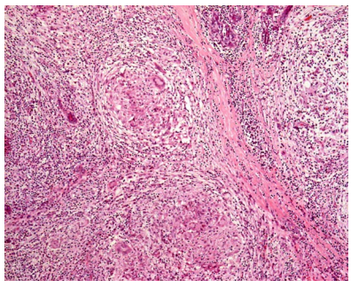
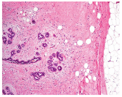
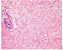
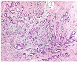
Sclerosing adenosis is a variant of breast pro- liferation. This is an important entity to recog- nize, as clinically it is characterized by an irregular hard mass that is fixed to the adjacent structures, and by imaging, it also shows significant architectural distortion, rendering this indistinguishable from carcinoma. Histologically, there is proliferation of epithelial and myoepi- thelial cells in the small ducts and ductules, and these are present within a densely fibrotic stroma. At times, the dense fibrosis or sclerosis will cause architectural distortion and compression of these ductal structures, giving rise to an infiltrative pattern (Fig. 2.4). Careful histologic examination to establish the benign nature of the epithelial cells, as well as to identify the pres- ence of an intact or attenuated myoepithelial celllayer, with the judicious use of immunohis- tochemistry for identification of the latter, helps to establish the diagnosis.
Other variants include blunt duct adenosis (columnar cell changes), microglandular adenosis, apocrine adenosis, and nodular adenosis. These will be discussed in greater details in Chap. 6.
Figure 2.4: Sclerosing adenosis with fibrosis of the intralobu- lar stroma resulting incompression of the epithelial structures to give an pseudoinfiltrative growth pattern.
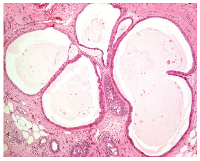
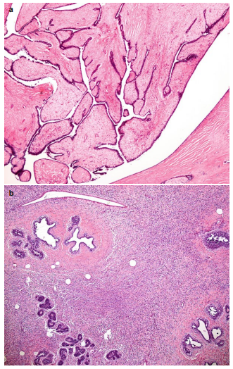
Fibroadenomas are probably the most com- mon benign breast tumor, presenting as solitary painless, mobile, and well-defined nodules. Multiple lesions are less frequent. Use of the immunosuppressant cyclosporine in transplant patients has resulted in an increased risk of fibroadenoma development. Macroscopically, fibroadenoma is ovoid, rubbery, and well cir- cumscribed; the cut surface is grayish and may be lobulated. Microscopically, it shows a mixed epithelial and stromal proliferation, giving rise to the pericanalicular and intracanalicular pat- terns, with the former formed by stromal prolif- eration around the ducts and the latter formed by compression of the ductal elements by the pro- liferating stromal component into slit-like spaces (Fig. 2.5). These patterns have little prognostic significance. Occasionally stromal giant cells, myxoid changes, dystrophic calcifications, and other mesenchymal metaplasia have been described. Complex fibroadenomas are those that show cysts larger than 3 mm, sclerosing adeno- sis, epithelial calcifications, or papillary apo- crine changes, and this group of fibroadenomasshows slightly higher (1.6×) cancer risk com- pared to the usual fibroadenomas. Fibroadenomas are benign, and most do not recur after surgical excision. Hamartoma may present as a soft palpable mass or as breast asymmetry, and is usually round to oval and lobulated. Histologically, it shows ducts, lobules, interlobular fibrosis, smooth mus- cle, and adipose tissue in varying proportions (Tse et al. 2002). This is a benign tumor and rarely recurs (Fig. 2.6).
Figure 2.5: Fibroadenoma showing proliferation and expansion of the stroma that is of usually low cellularity, and the ductal element sometimes forms an intracanalicular pattern with a “leaflike” pattern.
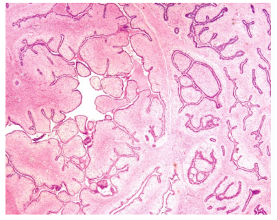
Figure 2.6: Hamartoma showing a rounded border, with “encapsulated”adipocytes within a fibrotic stroma. There is also intralobular fibrosis.
Diabetic mastopathy is an inflammatory disor- der of the breast characterized by a perilobular and perivascular lymphocytic infiltrate.
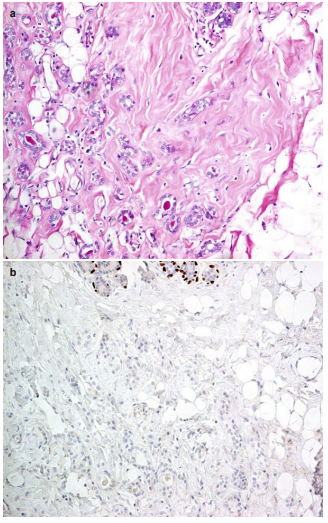
It is usually presents as a mass and is most common in women aged 25–60 years. It is characteristically associ- ated with long-standinginsulin-dependent diabetes mellitus and sometimes other autoim- mune diseases (also known as sclerosing lympho- cytic lobulitis). The radiologic features are variable, and mammography shows a dense parenchymal pattern with no specific mass, but sometimes, there is an asymmetric density and occasionally a cir- cumscribed mass. Ultrasound often shows a hypoechoic mass. The characteristic histological features are the presence of circumscribed aggre- gates of lymphocytes, with some plasma cells, around lobules, ducts, and vessels. The interlobu- lar stroma is fibrotic with plump epithelioid fibroblasts. In the stroma, keloidal fibrosis is seen. Phyllodes tumor is uncommon fibroepithelial neoplasm that resembles fibroadenoma grossly. Patients with phyllodes tumors usually are older than patients with fibroadenomas, and there may be a history of a rapidly growing mass. Multifocality and bilateriality are rare. Imaging may show a rounded, well-defined mass with clefts or compressed cystic spaces, and occasionally coarse calcifications are noted. Macroscopically, phyllodes tumor is a well-circumscribed, firm, bulging mass, and the cut section shows a fleshy mass with curved spaces resembling leaves or leaf buds. There may be hemorrhage or necrosis. Microscopically, phyllodes tumor shows a prom- inent intracanalicular growth pattern with leaflike patterns projecting into lumens. The epithelial component is usually benign, with an intact myoepithelial cell layer separating it from the stroma. The stroma is of higher cellularity than fibroadenoma and may show geographic varia- tion within the lesion. These stromal cells are bland looking, with scanty mitotic figures (Fig. 2.7a, b). Within the tumor, stromal areas of low cellularity, hyalinization, or myxoid changes are not uncommonly seen. Some examples of phyllodes tumors show stromal cell atypia and pleomorphism, increased mitotic activity, stromal overgrowth, and infiltrating margins, and these are considered phyllodes tumors of border- line or frank malignancy. Malignant phyllodes tumor behaves like a sarcoma rather than carci- noma and is further discussed in the section on sarcomas. Most of the phyllodes tumors are benign based on microscopy. Outcome of phyl- lodes tumor is dependent on the histologic grade. Benign phyllodes tumor may rarely recur but does not metastasize.
Figure 2.7 (a): Benign phyllodes tumor showing a fronded appearance, with variable stromal cellularity,and a benign epithelial lining on the surface. (b) Benign phyllodes tumor at higher magnification showing areas of stromal expansion with moderately cellular stroma.
Malignant phyllodes tumors including those of borderline or frank malignancy may both recur or metastasize, more commonly with the latter group (Tse and Tan 2005).
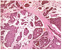
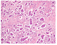
Papillomas can be divided into solitary or mul- tiple. Solitary papilloma is usually located beneath the nipple, whereas the multiple papillomas are more peripherally located. The former is more likely to present as nipple discharge and the latter is usually asymptomatic. Mammography may show a mass in solitary papilloma but multiple nodules or calcifications in peripheral papillomas. Ultrasound may highlight the cystic component particularly in the palpable examples. Micro- scopically, papillomas are characterized by an arborescent growth derived from the wall of a dila- ted duct, and these stromal tissue fibrovascular cores are lined by epithelial cells, together with an intervening layer of myoepithelial cells (Fig. 2.8a). The epithelial cells are benign, but apocrine or squamous metaplasia may be seen. Frequently, there is superimposed epithelial hyperplasia particularly of the florid type, and this will lead to complex architecture and obliteration of the ductal lumen (Fig. 2.8b). The fibrovascular cores may also show sclerotic changes resulting in compression and entrapment of the benign glandular component to yield a pseudo-invasive pattern. Demonstration of a layer of residual myo- epithelial cells as well as the absence of cellular atypia of these entrapped epithelial cells would help to differentiate this phenomenon from malig- nancy (Mulligan and O’Malley 2007). On the whole, papillomas are benign but are associated with slightly increased risk for cancer, more for the multiple papillomas than for the solitary papilloma (Lewis et al. 2006).
Figure 2.8(a): A benign papilloma showing a network of fibrovascular cores lined on the outside by benign ductal epithelial cells. The lesion is rounded and is present within a large ductal space. (b): A benign papilloma with a solid area of epithelial hyperplasia. The hyperplastic epithelium shows spindled nuclei with nuclear streaming, a feature characteristic of florid epithelial hyperplasia.
Epithelial Proliferative Lesions
Microglandular adenosis is an uncommon form of glandular proliferation. The proliferating tubules are lined by a single layer of epithelial cells devoid of a myoepithelial cell layer (Fig. 2.9a, b). Within the lesions, the epithelial cells form irregular tubules, and these epithelial cells are cytologically benign. Although myoepi- thelial cells are absent, these epithelial tubules have been shown to have an intact basement membrane. Microglandular adenosis has previ- ously been considered benign (Millis and Eusebi 1995), however, recent studies showed that it might be a nonobligate precursor of triple- negative breast cancer.
Figure 2.9(a): Microglandular adenosis showing permeation of the breast stromal tissue by small tubules formed by bland-looking epithelial cells with rounded lumens. Eosinophilic material may be seen within the lumens.
(b) Immunohistochemical staining of microglandular adenosis for the myoepithelial marker p63 shows a lack of myoepithelial lining around the tubules (terminal duct lobular units intact layer of myoepithelial).
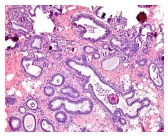
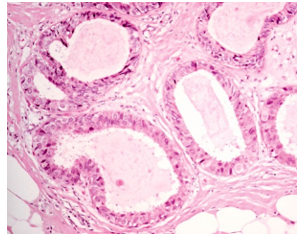
Columnar cell changes and columnar cell hyperplasia represent another spectrum of breast epithelial changes. These are characteristically non-palpable and are frequently detected either as incidental findings or by mammography for the associated calcifications. Microscopically, these lesions show well-maintained lobular archi- tecture, with dilated acini. The luminal cells are columnar, and they show apical snouting. Flocculent material is seen within the dilated lumens, and these are frequently associated with calcifications (Fig. 2.10).
Figure 2.10: Columnar cell changes showing dilatation of the acini with the cells demonstrating columnar morphology, with apical cytoplasmic snouts and the presence of flocculent material and calcifications within the lumens.
The lesion is called columnar cell change when there are only one to two layers of epithelial cells and termed colum- nar cell hyperplasia when there are more than two layers. The term flat epithelial atypia (FEA) is used when the epithelial cells show mild cyto- logical atypia (Fig. 2.11). Some studies suggested that at least some FEA may represent precursors for low-grade ductal carcinoma in situ (DCIS), but the risk of local recurrence or progression to invasion is very low (Schnitt 2003).
Figure 2.11: Flat epithelial atypia showing dilated acini containing secretory material. The spaces are lined by one to two layers of columnar epithelial cells with apical snouts and monomorphic nuclei. The nuclei are similar in appearance to those of ADH or low-grade DCIS.
Epithelial hyperplasia is the designation for proliferation of epithelial cells within preexisting ductal or lobular spaces. In general, epithelial
hyperplasia can be divided into usual hyperplasia or atypical hyperplasia. Interestingly, usual hyperplasia has only been described with ductal morphology (intraduct hyperplasia, epitheliosis, hyperplasia of the usual type, or usual duct hyper- plasia), whereas for atypical epithelial hyperpla- sia, both ductal and lobular morphologies have been described (atypical ductal hyperplasia (ADH) or atypical lobular hyperplasia (ALH)).
Usual ductal hyperplasia may be of variable size and extent, and although the morphology can be somewhat similar to low-grade DCIS, such a concept of progression is not generally accepted. The changes in usual ductal hyperplasia range from mild epithelial hyperplasia, in which the number of epithelial cell layers increased to two to four to florid epithelial hyperplasia, in which there are solid epithelial clusters within and oblit- erating ductal lumens, with formation of slit-like irregular peripherally located secondary lumens. The epithelial cells show mostly oval nuclei with irregular and streaming arrangement (Fig. 2.12). Apocrine metaplasia may be present.
Figure 2.12: Florid epithelial hyperplasia showing distension of ductal space by an epithelial proliferation,with the epithelial cells showing nuclear streaming and forming irregular and slit-like secondary lumens.
ADH, when properly defined using stringent cri- teria (Page and Rogers 1992), carries a significant cancer risk. The differentiation of atypical ductal hyperplasia from low-grade DCIS remains contro- versial and poorly standardized, with some authors reporting involvement of less than two glandular spaces or less than 2 mm being indicative of atypi- cal ductal hyperplasia (Page and Rogers 1992;Tavassoli and Norris 1990). On the whole, the his- tologic features of ADH may be viewed as a smaller version of low-grade DCIS (Fig. 2.13). Where the cutoff is drawn is still unsettled although the WHO consensus is to use 2 mm as the threshold (Lakhani et al. 2012).
Malignant Breast Tumors
Carcinoma In Situ
With the increasing use of mammographic screening, ductal carcinoma in situ (DCIS) is increasingly diagnosed as a non-palpable lesion. The traditional classification based on the archi- tectural pattern is now out of favor. Newer classifications/grading always use nuclear grade as one of the defining features for DCIS. Other histologic features being used are necrosis and the presence of tumor cell polarization – the organization of the nuclei around lumens within the tumor, resulting in rosette or cribriform structures (Silverstein et al. 1995; Holland and Hendricks 1994).
High-grade DCIS is easily differentiated from benign lesions, with the highly pleomor- phic tumor cells present within the enlarged ducts associated with central comedo necrosis. The associated calcifications within the necrotic debris produce a characteristic casting or branching pattern in mammography (Fig. 2.14). The ducts may be so distended that aggregation of these ducts may become palpable. Low-grade DCIS, on the other hand, shows monotonous and uniform tumor nuclei that may sometimes be difficult to distinguish from benign epithe- lial hyperplasia. Common histologic patterns include cribriform, with geometric punched out lumens within the tumor cell proliferation, or micropapillary with the proliferating tumor cells extending into the lumen without fibrovascular stalks (Fig. 2.15). The solid pattern is less com- mon. Secretions may be seen within the ductal lumen and should not be confused with necrosis. Classifications, if present, are associated with the secretions and are usually smaller, rounded, and psammomatous. Intermediate-grade DCIS usually shows features in between high- and low-grade lesions.
Lobular carcinoma in situ is sometimes grouped with ALH under the umbrella term of lobular neoplasia. In both cases, the lobular archi- tecture is essentially preserved, but individual acini are enlarged and distended with obliterated lumens. The neoplastic cells are small and uni- form, smaller than the ductal lesions, with higher nuclear cytoplasmic ratio, mild nuclear pleomor- phism, rare mitoses, and occasional cytoplasmic vacuoles (Fig. 2.16). Calcifications are much less frequent than ductal lesions. The differentiating features between ALH and lobular carcinoma in situ are not well defined, with the former showing generally less lobular distension and lesser extent of involvement, usually limited to part of a ductal-lobular unit.
Figure 2.13: Atypical ductal hyperplasia showing proliferation of a monomor- phic population of epithelial cells forming a cribriform structure. The cellular monotomy and architectural pattern are similar to those seen in low-grade DCIS.
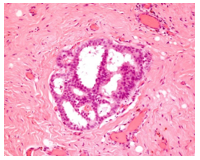
Figure 2.14: High-grade ductal carcinoma in situ with highly pleomorphic tumor cells present within and distending the duct space, associated with central comedo necrosis.
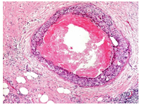
Figure 2.15: Low-grade ductal carcinoma in situ with monotonous tumor cells distending ductal spaces and forming cribriform structures with geometric lumens.
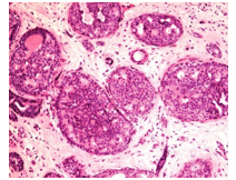
Figure 2.16: Lobular neoplasia showing distension of the acini by a uniform population of small round cells with bland nuclear features. The growth pattern is solid.
PapillaryCarcinoma
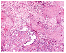
Papillary carcinomas are uncommon malignant lesions, representing several different morphologi- cal entities, all possessing a common papillary archi- tecture, characterized by epithelial proliferation overlying elaborate fibrovascular cores. Papilloma with DCIS and intracystic papillary carcinoma are typical examples of papillary lesions with malig- nancy. Papilloma with DCIS usually occurs when there is a focus of atypical epithelial proliferation within an otherwise benign papilloma. This phenomenon is not unusual, and the atypical epithelial focus usually possesses the usual histo- morphology of uncomplicated ADH or low-grade DCIS (Fig. 2.17). The differentiation between pap- illoma with ADH or papilloma with DCIS is arbi- trary, with a size criterion of 3 mm being used. If the atypical focus is larger than 3 mm, the lesion should be termed as papilloma with DCIS, and when the focus is 3 mm or less, it should be termed papilloma with ADH (Page et al. 1996).
Figure 2.17: Papilloma involved by atypical ductal hyperplasia. There are areas of atypical ductal hyperplasia separating the papillary fibrovascular stromal tissue cores. The atypical ductal hyperplasia shows typical morphology with rounded cells forming geometric structures.
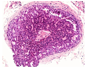
Intracystic or encapsulated papillary carcinoma is rare, usually occurring in older women, and may present as a breast mass. Microscopically, it is predominantly papillary but may also exhibit cri- briform or micropapillary patterns as minor com- ponents. The characteristic feature of this tumor is the essentially absence of a complete layer of myoepithelial cells on the outside and the deli- cate nature of the papillary fronds (Collins et al. 2006) (Fig. 2.18). Intracystic papillary carcinoma has a good prognosis, having better outcome than mixed intracystic papillary/nonpapillary tumors (Carter et al. 1983; Lefkowitz et al. 1994). Most recommend a treatment protocol more akin to that of an in situ disease.
Figure 2.18: Intracystic papillary carcinoma showing delicate fibrovascular cores within a rounded tumor, with the absence of an outer myoepithelial cell layer.
Invasive Carcinoma
Invasive breast carcinomas are classified based on their histological features, and this classification also reflects their clinical behavior. Incidence increases with patient’s age, with family history being one of the most common risk factors. Clinically, it presents as an ill-defined mass, sometimes adherent to the skin or underlying muscle.
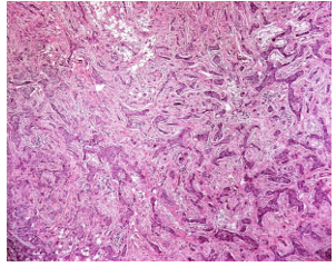
Among the tumor types, the most common is invasive ductal carcinoma, not otherwise specified (IDC, NOS). The tumor is of varying size and may be associated with calcification. It is usually firm, fibrotic, or stellate. The histomorphology of the tumor is also highly variable, ranging from a low-grade tumor showing mildly pleomorphic tumor cells arranged in tubules with little mitoticactivity to a high-grade tumor showing highly pleomorphic tumor morphology, with the tumor cells arranged in solid sheets and groups, show- ing brisk mitotic activity and abundant tumor necrosis (Fig. 2.19). Most IDCs are graded based on three microscopic features, namely, the degree of tubule formation, nuclear pleomorphism, and mitotic count. The tumor cells in higher-grade cancer are usually arranged in sheets or are dis- cohesive, showing very little tubule formation, whereas lower-grade cancer shows significant tubule formation generally. Nuclear morphol- ogy is also evaluated in terms of variation in nuclear size, regularity of the nuclear border, hyperchromasia, and prominence of nucleolus. Mitoses are usually counted per 10 high-power microscopic fields, taking into account the sizeof the microscopic high power field. A combi- nation score on these three components reflects tumor grade and prognosis. Furthermore, hor- mone receptor assessment is also mandatory in tumor evaluation. ER, PR, and HER2 protein expression are routinely performed by immuno- histochemistry, and the results carry prognostic and predictive significance. Tumors expressing hormone receptors have better prognosis and are highly responsive to hormonal therapy, whereas tumors expressing HER2/neu have poor progno- sis but may respond to specific anti-HER2/neu targeted therapy.
Figure 2.19: Infiltrating duct carcinoma showing irregular groups of malignant cells invading a desmoplastic stroma. The normal ductal-lobular architecture is obliterated.
Invasive lobular carcinoma represents 5–10 % of the invasive breast tumors. It is usually seen as an irregular thickening and with a lower density in mammography and less likely to be associated with calcification. The tumor can be relatively small and possesses poorly defined edges. The tumor cells are small and subtle, containing small amounts of cytoplasm. There is no definite configuration except the formation of cords or single file pattern (Fig. 2.20). The tumor cells usually show loss of E-cadherin, an epithelial cell adhesion molecule, and this can be demonstrated immunohistochemically, a fact that can be uti- lized in diagnosis. The loss of E-cadherin expres- sion in ILC seems to be associated with evidence of impaired integrity of the E-cadherin catenin membrane complex. Whether invasive lobular carcinoma has a worse prognosis than invasive ductal carcinoma is still controversial, with numerous studies presenting contrasting results. However, contralateral invasive carcinoma is reported to be more frequent in patients with a history of invasive lobular carcinoma.
Figure 2.20: Infiltrating lobular carcinoma showing poorly cohesive small round tumor cells arranged in single files within a fibrotic stroma.
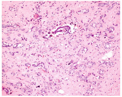
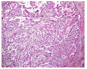
Tubular carcinoma is a rare breast tumor, accounting for less than 3 % of all the tumor types. Most of these tumors are detected via the screening program with a characteristic mammo- graphic appearance of an irregular spiculated mass usually lacking calcification. The tumor is usually small and composed of angulated tubules embedded in a desmoplastic stroma (Fig. 2.21). This is one of the well-differentiated tumors that are commonly misdiagnosed as benign at fine needle aspiration or biopsy. The absence of myo- epithelial cells helps to confirm a diagnosis of tubular carcinoma. Lymph node metastasis is rare and 5-year survival rate of patients with this tumor type is more than 90 %.
Figure 2.21: Tubular carcinoma showing bland-looking tumor cells arranged in tubules that possess widely patent lumens. Myoepithelial cells are absent, and the malignant tubules are arranged in an irregular stellate pattern. The intervening stroma is densely fibrotic.
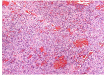
Mucinous carcinoma occurs commonly in postmenopausal women, presenting at the clinic with a palpable mass. The tumor is usually circumscribed and associated with radiologic microcalcification. Grossly, a large gelatinous appearance is appreciated. Microscopically, the tumor cells are usually in tubules, clusters, and small sheets floating in pools of mucin. They have a characteristic eosinophilic cytoplasm with low nuclear and mitotic grade. The amount of tumor cells varies, and hypercellular tumors may sometimes show neuroendocrine differentiation (Fig. 2.22). Furthermore, neuroendocrine dif- ferentiation in mucinous carcinoma is associated with favorable histologic and immunohistochem- ical parameters.
Figure 2.22: Mucinous carcinoma showing large sheets of low-grade malignant cells present within pools of accumulated extracellular mucinous material.
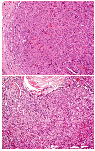
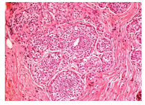
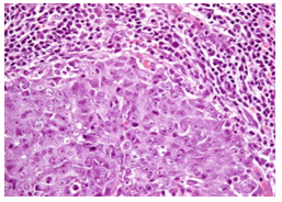
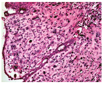
The presence of the malignant cells is a prerequisite in the diagnosis in order to distinguish it from benign mucocele-like lesions. Generally, mucinous carcinoma confers good prognosis, mostly diagnosed as grade I or II with 5-year survival of 90 %. Carcinoma with medullary features has a dis- tinctive morphological appearance. Grossly the tumor is a sharply circumscribed mass with soft and fleshy consistency which may be confused with fibroadenoma, having a gray and solid cut surface. The tumor cells are seen in a syncy- tial growth pattern, with pushing margins and prominent lymphoplasmacytic infiltrates in the perimeter (Fig. 2.23).
Figure 2.23: Carcinoma with medullary features showing a rounded margin and an intense lymphocytic infiltrate at the periphery. The tumor cells form a sheetlike pattern with high degree of nuclear pleomorphism and brisk mitotic activity.
Morphological appear- ance of the cells is high grade and usually with more than 20 mitotic figures per 10 high-power fields. Furthermore, this histologic type is also seen among patients with BRCA1-mutation- related tumors. However, it does not translate that all BRCA1-related tumors should be called medullary like. Nevertheless, these morpho- logical features should prompt the search for a family history of a genetic predisposition, espe- cially when a young patient is involved. BRCA1 is an independent predictor of disease-free interval, and its alteration may play a role in the development and progression of breast cancer (Rakha et al. 2008). The tumor cells are gener- ally negative with ER, PR, and HER2 stains.
Metaplastic carcinoma encompasses a mixed range of uncommon cancers and has been used by pathologists to describe breast carcinomas of mixed epithelial and mesenchymal appearance. Overall, tumors in this group represent less than 1 % of all invasive breast tumors. These tumors are usually larger compared to other types, with ill-defined shapes and surface. Malignant squamous cells and glands are often the histo- logical components encountered mixed with spindle cells (Fig. 2.24).
Figure 2.24: Metaplastic carcinoma showing mixed squamous cell carcinoma and ductal carcinoma components. The intervening areas show malignant spindle cell proliferation.
They are further catego- rized based on the predominance of the epithelial and/or mesenchymal cells. Generally, the prog- nosis is poor, showing increased incidence of recurrence and metastasis. Invasive micropapillary carcinoma accounts for approximately 2 % of all invasive breast car- cinomas and appears associated with a poor prog- nosis. The tumor cells are seen as tubular nests surrounded by clear spaces which may be related to an artifactual tissue shrinkage. The tubules lack true fibrovascular cores and exhibit reverse polarity of cells (luminal markers on the periphery of islands) (Fig. 2.25). This tumor is predomi- nantly of histologic grade III with a higher inci- dence of lymphatic invasion and lymph node metastasis.
The eosinophilic and granular cytoplasm of apocrine carcinoma is its distinguishing feature (Fig. 2.26). This is an uncommon tumor that exhib- its the usual tubular or solid arrangement of the tumor cells. Similarly, the size, grade, and lymph node stage are the same with that of invasive ductal carcinoma. The tumor cells stain positively with androgen receptor (AR) and GCDFP-15.
Figure 2.25: Invasive micropapillary carcinoma showing rounded groups of tumor cells with a peripheral clear rim.
Inflammatory carcinoma is a clinical descrip- tion of the tumor which on presentation shows skin redness, warmth, and an edematous appearance. This tumor mimics an infection and may transiently respond to medications; thus, clinical caution is important. The prognosis is usually poor and usu- ally associated with dermal lymphatic permeation.
Figure 2.26: Apocrine carcinoma showing tumor cells with the characteristic appearance of abundant eosinophilic and granular cytoplasm.
Angiosarcoma of the breast is tumor arising from the blood vessels and is grossly seen as a poorly defined mass with hemorrhage. When an old patient is involved, it is most of the time sec- ondary to chronic lymphedema or radiotherapy of prior breast cancer. Microscopically, vasofor- mative cells with endothelial tufting with atypia are seen. Necrosis and hemorrhage are common in aggressive tumors (Fig. 2.27).
Figure 2.27: Angiosarcoma showing malignant spindle cells with moderate nuclear pleomorphism forming vascular channels of varying sizes and shapes. Some of these lumens are blood filled.
Malignant phyllodes tumor is distinguished from the benign tumor based on the microscopic features of the stromal element. Stromal hyper- cellularity, overgrowth, atypia, and mitotic activity of 10 or more per ten high-power fields are the features seen in malignant phyllodes which will also show an infiltrative margin (Fig. 2.28). Management is usually difficult, and most surgeons will perform mastectomy once a malig- nant diagnosis is made. Incidence of recurrence and metastasis is also increased.
Figure 2.28: Malignant phyllodes tumor showing highly pleomorphic malignant stromal cells, with bizarre cells adjacent to benign ductal epithelium.Atypical mitotic figures are seen within the malignant stromal cells.
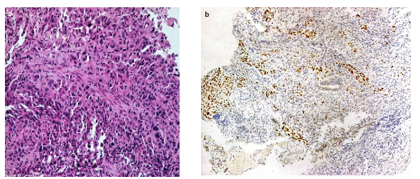
When entertaining metastatic tumors in the breast, a full knowledge of the clinical history is important to avoid misdiagnosis. The tumor will commonly present as small nodules which on microscopy will reveal a peculiar architecture that is different from the common breast histo-logic types. The common metastatic tumors in the breast are lung carcinoma and malignant mel- anoma. The tumor cells will usually be high-grade or anaplastic, making it more difficult to decipher the similarity within its origin. Presence of an in situ carcinoma may give a hint of a primary breast tumor, but its absence will also not rule it out. The use of special stains to identify tumor origin is most helpful, such as TTF-1 in detecting metas- tasis from lung adenocarcinoma (Fig. 2.29a, b).
Figure 2.29: High-grade malignant cells presenting as a tumor nodule in the breast. The tumor cells show hyperchromatic nuclei and moderate amounts of cytoplasm. (b): The same tumor showing positivity for TTF1, confirming a metastasis from a lung primary.
References
- Akcan A, Akyildiz H, Deneme MA et al (2006)Granulomatous lobular mastitis: a complex diagnosticand therapeutic problem. World J Surg 30:1403–1409.
- BakarisS,YukselM,CiragilPetal(2006)Granulomatousmastitisincludingbreasttuberculosisandidiopathiclobular granulomatous mastitis. Can J Surg 49: 427–430.
- Carter D, Orr SL, Merino MJ (1983) Intracystic papillarycarcinomaofthebreastaftermastectomy,radiotherapyorexcisionalbiopsyalone.Cancer52: 14–19.
- CollinsLC,CarloVP,HwangHetal(2006)Intracysticpapillarycarcinomaofthebreast:are-evaluationusing a panel of myoepithelial markers. Am J Surg Pathol30: 1002–1007.
- HollandR,HendricksJ(1994)Microcalcificationsassociated with ductal carcinoma in situ: mammographic-pathologiccorrelations.SeminDiagnPathol11:181–192.
- LefkowitzM,LefkowitzW,WargotzES(1994)Intraductal(intra cystic) papillary carcinoma of the breast and itsvariants: a clinicopathologic study of 77 cases. HumPathol25: 802–809.
- Lewis JT, Hartmann LC, Vierkant RA et al (2006) Ananalysis of breast cancer risk in women with single,multiple, and atypical papilloma. Am J Surg Pathol30: 665–672.
- Millis RR, Eusebi V (1995) Microglandularadenosis ofthebreast. Adv Anat Pathol 2: 10–18.
- Mulligan AM, O’Malley FP (2007) Papillary lesions ofthebreast:areview.AdvAnatPathol14: 108–119.
- Page DL, Rogers LW (1992) Combined histologic andcytologiccriteriaforthediagnosisofmammaryatypicalductalhyperplasia.HumPathol23:1095–1097.
- PageDL,SalhanyKE,JensenRAetal(1996)Subsequentbreast carcinoma risk after biopsy with atypia in abreast papilloma. Cancer 78: 258–266.
- RakhaEA,El-SheikhSE,KandilMAetal(2008)Expression of BRCA1 protein in breast cancer and itsprognosticsignificance.HumPathol39: 857–865.
- Schnitt SJ (2003) The diagnosis and management of pre-invasivebreastdisease:flatepithelialatypiaclassification,pathologic features and clinical significance. BreastCancerRes5: 263–268.
- SilversteinMJ,PollerDN,WaismanJRetal(1995)Prognostic classification of breast ductal carcinoma insitu. Lancet 345: 1154–1157.
- Simpson JF, Schnitt SJ, Visscher D et al (2012) Atypicalductal hyperplasia. In: Lakhani SR, Ellis IO, SchnittSJ, et al (eds) WHO Classification of Tumours of theBreast.IARC Press, Lyon. p.88
- Tavassoli FA, Norris HJ (1990) A comparison of theresults of long term follow up for atypical intraductalhyperplasia and intraductal hyperplasia of the breast.Cancer 65: 518–529.
- TseGM,TanPH(2005)Recentadvancesinthepathologyof fibroepithelial tumours of the breast. Curr DiagnPathol11: 426–434.
- Tse GM, Law BK, Ma TK et al (2002) Hamartoma of thebreast:aclinicopathologicalreview.JClinPathol55: 951–954.