T Cell Lymphoma associated with Langerhans Cell Histocytosis in Sudanese Patient
Adil HH Bashir1*, Lamyaa AM Elhassan5, Abdel Khalig Muddathir6, Khalid O. Alfarouk1, Ahmed M. Elhassan1,2
1 Institute of Endemic Diseases, University of Khartoum, Khartoum, Sudan.
2 Faculty of Medicine, University of Bahri, Khartoum North, Sudan.
3 Uneizah Pharmacy College, Qassim University, Alqassim, KSA.
4 Faculty of Pharmacy, Omdurman Islamic University, Khartoum, Sudan.
5 School of Medicine, Ahfad University for Women, Omdurman, Sudan 6Faculty of Pharmacy, University of Khartoum, Khartoum, Sudan.
*Corresponding Author: Adil HH Bashir, Institute of Endemic Diseases, University of Khartoum, Khartoum, Sudan, Tel: +24912966733 / +249960311336; Fax: +249960311336; E-mail: derma55@yahoo.com
Citation: Adil HH Bashir, Lamyaa AM Elhassan, Abdel Khalig Muddathir, Khalid O. Alfarouk, Ahmed M. Elhassan, et al. (2023) T Cell Lymphoma associated with Langerhans Cell Histocytosis in Sudanese Patient. J Dermatol & Ther 3: 117.
Received: April 11, 2023; Accepted: April 18, 2023; Published: April 22, 2023.
Copyright: © 2023 Adil HH Bashir, et al. This is an open-access article distributed under the terms of the Creative Commons Attribution License, which permits unrestricted use, distribution, and reproduction in any medium, provided the original author and source are credited.
Abstract
Cutaneous T-cell lymphomas (CTCLs) are the largest group of cutaneous lymphomas, representing 65% of all cutaneous lymphomas. Dendritic cells (DCs) are potent antigen- presenting cells that help orchestrate the innate and adaptive immune systems to induce tolerance and immunity. However, DCs may perform a dual role in the pathogenesis of CTCLs. Immature DCs (Langerhans cells) could promote the survival of malignant T cells. Further understanding of DCs and their role in CTCLs can help us to uncover the pathogenesis of this disease and to further explore the therapeutic uses of DCs. We reported rare case of adult T cell lymphoma associated with Langerhans cells occurred in a 43-year-old Sudanese adult, presented as universally dry scaly skin with large 5X6 cm fungating and discharging ulcer at vertex and small one at nape, puffy face and swollen frontal area for 5 years duration. The case was diagnosed and confirmed histopathologically, considered to be the first case been reported in Sudan.
Keywords
T cell lymphoma, Langerhans cell Histocytosis, Sudan.
Introduction
Cutaneous T-cell lymphomas (CTCLs) are the largest group of cutaneous lymphomas, representing 65% of all cutaneous lymphomas. The World Health Organization (WHO)/European Organization for Research and Treatment of Cancer (EORTC) classification (WHO-EORTC classification) is used to categorize CTCLs. [1, 2, 3] However, a substantial subset of T-cell primary cutaneous lymphomas remains that cannot be classified beyond the unspecified peripheral T- cell category, some of which may have an aggressive course.[4]
Blastic NK-cell Lymphoma is a very rare cancer, affecting only a few people (usually adults) each year. This lymphoma was previously thought to arise from a T- or NK-cell. However, newer studies indicate that it may arise from another type of white blood cell called a plasma, or dendritic cell. This lymphoma is fast-growing and can be difficult to treat. It can arise anywhere in the body. Dark red or purple skin lesions are a common feature. Dendritic cells (DCs) are potent antigen- presenting cells that help orchestrate the innate and adaptive immune systems to induce tolerance and immunity. However, DCs may perform a dual role in the pathogenesis of CTCLs. Immature DCs (Langerhans cells) could promote the survival of malignant T cells. Further understanding of DCs and their role in CTCLs can help us to uncover the pathogenesis of this disease and to further explore the therapeutic uses of DCs.[5] Cutaneous T-cell lymphoma (CTCL) is a malignancy derived from a clonal population of mature, skin-homing lymphocytes. In the skin, the CTCL cells are associated with the Langerhans cells and respond to protumor cytokines.[6] Scabies, infection with Sarcoptes scabiei, is known to be predisposed to by poor body hygiene, environmental exposure, and systemic immunodeficiency. Notably reduced numbers of Langerhans cells. These findings suggest that the development of scabies may be predisposed to by local cutaneous immunodeficiency secondary to reduced numbers of Langerhans cells.[7]
Case Presentation
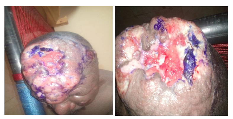
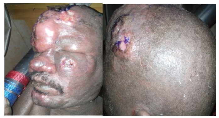
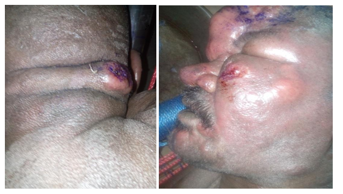
A male patient, single, 43 years old, laborer, descent from first degree relative parents, resident in Taief, Gaalei tribe, was referred to our clinic complaining of widespread dry scaly skin lesions with large 5X6 cm punch out discharging ulcer at vertex and small one at nape, puffy face and swollen frontal area. No regional lymph nodes are palpable. The condition was not associated with fever or other constitutional manifestations. The condition started 5 years ago with insidious onset and progressive course.
General examination: The general condition is well, not pale, not icteric, as well no palpable spleen, and liver. No palpable lymph nodes.
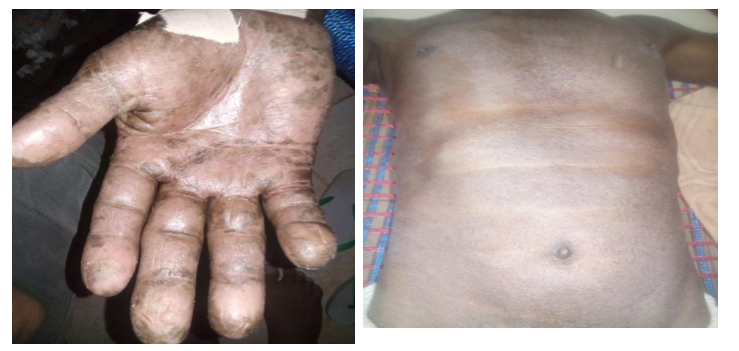
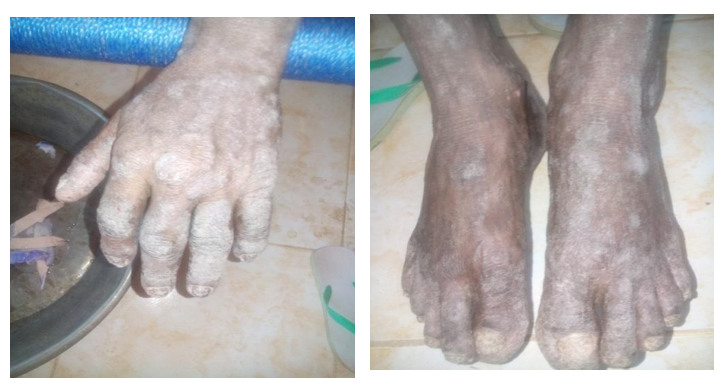
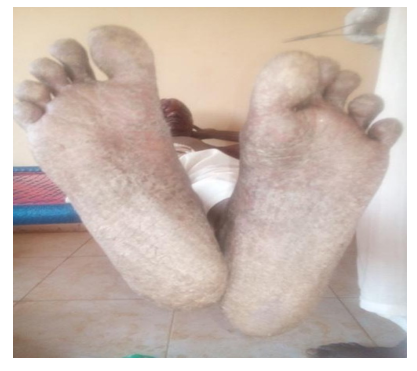
Dermatological examination: widespread dry scaly skin lesions with large 5X6 cm punch out, elevated margins, discharging, serosangionus ulcer at vertex and small one at nape. Skin is erythrodermic and scaly with Palmoplantar hyperkeratosis. No palpable regional lymph nodes.
Palms and soles: Hyperkeratotic lesions at both soles and palms.
Nails: Dystrophy has been noticed.
Ears: No Abnormality Detected.
Hair: No Abnormality Detected.
Oral cavity: No Abnormality Detected.
Figure 1: Punch out discharging ulcer at vertex.
Investigations Result
Date: 31/3/2015. Slide: # 429/H/015.
Sections show poorly formed nodules in the dermis consisting mainly of lymphocytes. The majority of the cells are CD3 but there are also less CD20 positive cells. BCL2 is mildly positive in one focus. There are many CD1a positive Langerhans cells.
Diagnosis: The appearance is highly suggestive of CD3 lymphoma. Some cases of cutaneous T cell lymphomas associated with Langerhans cells have been described.
Skull X-Ray (AP & LAT)
No bone density; No lytic lesions or periosteal reactions.
Normal skull sutures, no abnormal vascular markings.
There is a large scalp soft tissue ulcer. Investigations:
CRP Positive
Serum Albumin 2.4 Low
CRP Positive
Serum Albumin 2.4 Low
TLC 1.3Low
Serum Potassium 4.0
Low Serum Calcium 7.7 Low
Serum LDL 600 High
HCV Negative
HBsAg Negative
HIV Negative
Wound C&S Pseudomonas Aregenosa
Discussion
The patient presented here is unique and demonstrates a spectrum of diseases associated with CTCL and LCH. Blastic NK-cell Lymphoma is a very rare cancer, affecting only a few people (usually adults) each year. This lymphoma was previously thought to arise from a T- or NK-cell. However, newer studies indicate that it may arise from another type of white blood cell called a plasma, or dendritic cell. This lymphoma is fast- growing and can be difficult to treat. It can arise anywhere in the body. Dark red or purple skin lesions are a common feature.
Conclusions
Cutaneous T cell lymphomas associated with Langerhans cells may arise from another type of white blood cell as dendritic cell.
Acknowledgments
This work has been supported by Alfarouk Biomedical Research LLC.
References
1. Burg G, Kempf W (2005) Etiology and pathogenesis of cutaneous lymphomas. In: Burg G, Kempf W. Cutaneous Lymphomas. London: Taylor & Francis.
2. Willemze R, Jaffe ES, Burg G, et al. (2005) WHO- EORTC classification for cutaneous lymphomas. Blood 105: 3768-85.
3. Burg G, Kempf W, Cozzio A, et al. (2006) Cutaneous malignant lymphomas: update 2006. J Dtsch Dermatol Ges 4: 914-33.
4. Khandesi Z, Ben-Arieh Y, Izhak OB, Epelbaum R, Dann EJ, et al. (2008) The applicability of the new WHO-EORTC classification of primary cutaneous lymphomas to a single referral center. Am J Dermatopathol 30: 37-44.
5. Ni X, Duvic M (2011) Dendritic cells and cutaneous T-cell lymphomas. G Ital Dermatol Venereol 146: 103-13.
6. Jaehyuk Choi, Francine Foss (2007) Cutaneous T-cell lymphoma: Biologic targets for therapy. Current Hematologic Malignancy Reports. 2: 272-277.
7. Douglas H McGregor, Qinghai Yang, Fang Fan, Robert L Talley, Margarita Topalovski et al. Scabies Associated with Radiation Therapy for Cutaneous T-Cell Lymphoma. Address correspondence to Douglas H. McGregor, MD, Pathology and Laboratory Medicine (DRC/113), Veterans Affairs Medical Center, 4801 Linwood Boulevard, Kansas City, MO 64128, USA.