Direct Evidence of Viral Infection and Mitochondrial Alterations in the Brain of Fetuses at High Risk for Schizophrenia
Mesa CS, CUBA1*
*Corresponding Author:Segundo Mesa Castillo, Psychiatric Hospital of Havana, Cuba, Tel: 537 444690; Fax: 537 444690
Citation: Mesa CS (2023) Direct Evidence of Viral Infection and Mitochondrial Alterations in the Brain of Fetuses at High Risk for Schizophrenia. Addict drug sensitiz 4: 126.
Received: June 21, 2023; Accepted: June 28, 2023; Published: July 01, 2023.
Copyright: © 2023 Segundo Mesa Castillo, et al. This is an open-access article distributed under the terms of the Creative Commons Attribution License, which permits unrestricted use, distribution, and reproduction in any medium, provided the original author and source are credited.
Introduction
Schizophrenia with 1% of prevalence constitutes one of the fundamental problems of public health in the world for its human, social and economic repercussion independently of the temporary or geographical context where it appears.
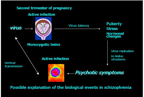
The neurodevelopmental hypothesis in the aetiology and physiopathology of schizophrenia is considered one of the most consistent at present. It is based on a series of evidences that guide toward an affectation in the critical period of the human being development due to pregnancy and delivery complications, particularly those with known or presumed impact on foetal neurological development, that result in increased risk for schizophrenia psychosis. Among the possible etiological candidates are viral infections. The minor physical and functional anomalies, manifesting as soft neurological sings, slight anatomical defects of the head, hair, eyes, mouth, hands and feet, as dermatoglyphic asymmetries, are due to some injury occurring during the first and more probable second trimester of foetal life, and are more common among patients with schizophrenia and in their unaffected siblings than in the general population. A virus acting in this important and critical stage of the development interacting or not with genetic factors can be responsible for the cascade of biological events that appear later on and could explain the period of relative stillness that exists between the birth and the appearance of the symptoms in the puberty that could be related to the reactivation of a latent viral infection. In the present work additional results are presented in an ultrastructural study carried out in samples of the left temporal lobe of foetuses aborted for medical reasons from schizophrenic mothers with strong familial antecedents of schizophrenia. The findings obtained are compatible with an active infection of the central nervous system by herpes simplex hominis type I [HSV1] virus during the second trimester of pregnancy. Until we
report evidences supporting the concept of virus-cell interaction in the neurodevelopmental hypothesis of schizophrenia had been indirect. Virus particles had never been demonstrated. The present results are the first direct evidence that demonstrate the presence of virus particles in the central nervous system of foetuses in the critical period of the second trimester of foetal development. The importance of this finding can have practical applications in the prevention of the illness keeping in mind its direct relation to the aetiology and physiopathology of schizophrenia. Among the measures of preventive character in pregnant women at risk of having a descendant of schizophrenia are the study of the amniotic fluid cells by electron microscopic techniques, and in consequence in case of being positive of viral infection, the recommendation of an early antiviral treatment or the voluntary interruption of pregnancy among other measures.
Results
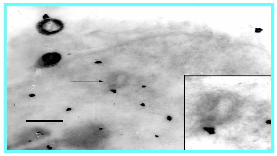
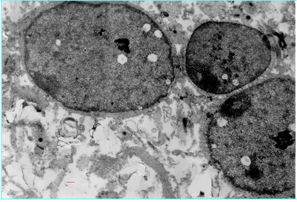
In four of five of the studied foetuses it was observed within the nucleus of neurons the presence of vacuoles with spherical empty particles of 100 nm occupying the center of an electron-lucid area [Fig.1]. The inclusions with particles appeared in number from 2 to 8 per nucleus, with great incidence in their appearance. The size and form of the particles coincides with the observations made of similar particles in the brain of post-mortem studies of adult schizophrenics [Fig. 2] and in animals experimentally inoculated with cerebrospinal fluid from schizophrenic patients [Fig. 3] using the same electron-microscopic techniques. The rest of the cells of the nervous system didn't present these particles. No particles were observed in a control study. A positive reaction to herpes simplex hominis type I antigen was observed when inmuno electromicroscopic techniques were used.
Figure 1: This finding is direct evidence obtained with a high-resolution power technique at cellular level in a stage of the foetal development not explored previously. and theoretically signal as vulnerable and explanatory of the later biological events in schizophrenia related to an aggression of the cell by environmental factors. An active viral infection of the central nervous system in this stage of the human development is demonstrated by the presence of immature viral particles. Bar: 200 nm.
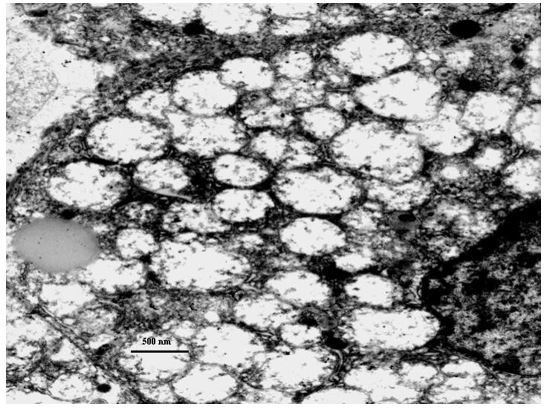
Figure 2: Young adult paranoid schizophrenic patient. Sample obtained from the left amygdala. Post mortem study Bar: 200 nm. Intranuclear particle.
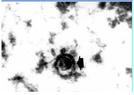
Figure 3: Chicken embryo inoculated with cerebrospinal fluid from a schizophrenic patient. Inset: viral particle labeled with peroxidase anti-herpes antibody. Intranuclear particles Bar 200 nm.
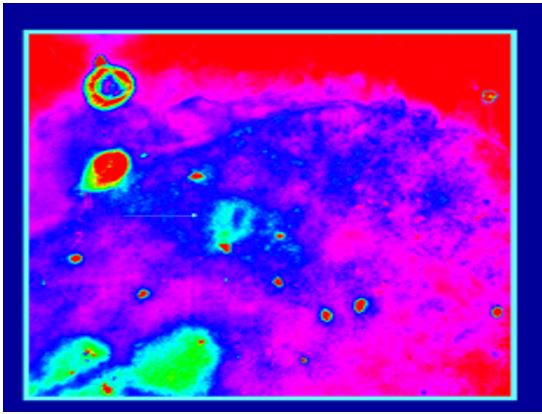
Digital analysis of image from figure 3.
Mitochondria Alterations
Although alternative explanations are possible, a most attractive possibility is that the infected neuron by HSV1 virus in foetal brain will serve as a source of latent virus and its later reactivation in limbic structures by environmental stressors would explain the recurrent character of schizophrenia. A vertical type transmission to offspring would restart the cycle of the illness in new generations.
Acknowledgements
Psychiatric Hospital of Havana; National Institute for Scientific Research of Cuba; partial financial support for this work from Faculty of Science, Málaga University, Prof. Sebastián Bruque.