Acute Hemorrhagic Leukoencephalitis- A Rare Medical Catastrophe!!!
Spandana Devi Tagaram*1, Sandeep Patil1, Shiji Chalipat1, Maya Patil1, G Karambelkar1, SR Agarkhedkar1
1Department of Pediatrics, Dr. D.Y. Patil Medical College, Hospital and Research Centre, Pune, Maharashtra, India
*Correspondence Author: Spandana Devi Tagaram, Department of Pediatrics, Dr. D.Y. Patil Medical College, Hospital and Research Centre, Pune, Maharashtra, India, Tel: +91 20 27805100; Fax: +91 020 27805217; E-mail: spandana.india@gmail.com
Citation:Spandana Devi Tagaram, Sandeep Patil, Shiji Chalipat, Maya Patil, G Karambelkar, et al. (2022) Acute Hemorrhagic Leukoencephalitis- A Rare Medical Catastrophe Pediatr Primary Physic 2: 109.
Copyright: © 2022 Spandana Devi Tagaram, et al. This is an open-access article distributed under the terms of the Creative Commons Attribution License, which permits unrestricted use, distribution, and reproduction in any medium, provided the original author and source are credited
Received date: March 28, 2022; Accepted date: April 06, 2022; Published date: April 11, 2022
Introduction
Acute hemorrhagic leukoencephalitis (AHLE) or Weston-Hurst’s syndrome is a hyper acute severe variant of acute disseminated encephalomyelitis(ADEM) with an incidence of 0.4 -0.5 per 1,00,000. It manifests with multiple neurologic symptoms, encephalopathy that may be progressive in nature, but has some recovery after treatment. It is characterized by a hemorrhagic inflammatory response in the central nervous system primarily affecting the white matter, myelin sheath often sparing the periventricular white matter. Most cases are autoimmune in origin and others have prior history of (h/o) viral illness and post vaccination status. It is a diagnostic challenge at its first attack.Specialised spin-ECHO Magnetic resonance Imaging (MRI) is the diagnostic .AHL usually has a very poor prognosis with high mortality. Treatment is unclear but high dose steroids/immunosuppresants may offer some benefit and promote better recovery although with a residual neurological deficit.
Case Report
Presenting a case of an 8-year-old female child was admitted with sudden onset of slurry speech, headache, urinary retention, vomiting, altered mental status followed by two weeks of febrile illness. She had H/O upper respiratory viral illness six weeks prior. Examination is positive for generalised hypotonia with brisk deep tendon reflexes and with extensor plantar response. Her Glassgow coma scale (GCS) was 8/15 on admission. Pupils were reactive to light and accomodation. Labs showed high inflammatory markers, normocytic normochromic anemia with benign renal, coagulation, hepatic function test panel. The initial impression was acute meningio-encephalitis, so broad spectrum antibiotics and antiviral drugs were commenced and lumbar puncture was performed after some period of stabilization. Viral encephalitis panel was negative along with Malaria, Dengue, Typhoid serologies. Lumbar puncture (LP): CSF protein-14.9, sugar -72mg/dl gross: clear, microscopy:5 nucleated cells(lymphocytes).
MRI: Bilaterally symmetrical areas of altered signal intensity involving ventrolateral thalamus, lateral mid brain, ventrolateral pons, and bilateral middle cerebellar peduncles appearing hyper intense on T2W1. T2W1, FLAIR sequences showing diffusion restriction appearing dark and foci of haemorrhages seen on SW1. Figure 1- 4
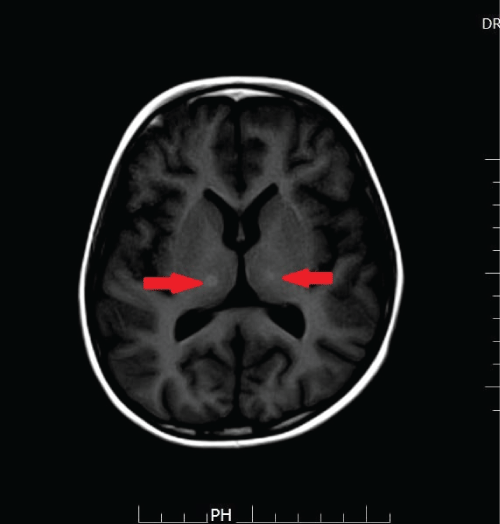
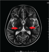
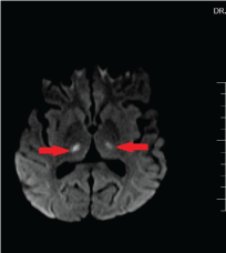
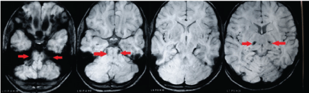
SW: SEQUENCE (Susceptability weighed): Bilateral foci of hemorrhages involving ventrolateral thalamus, lateral mid brain, ventrolateral pons, and bilateral middle cerebellar peduncles.
Patient is diagnosed to have acute hemorrhagic leukoencephalitis based on the clinical history and neuro imaging findings. She received three days of high dose pulse steroids followed by tapering of oral steroids for a total of 8 weeks. The patient showed a gradual improvement in the GCS score and her neurological symptoms resolved. She does have some residual neurological sequelae (spasticity of her extremities) at present.
Discussion
AHLE is a rare manifestation of ADEM. History is suggestive of 1) acute encephalopathy and neurological symptoms with h/o recent vaccine use or post viral phase and 2) MRI findings of demyelinating punctate hemorrhages in cerebral hemispheres. Differential diagnosis to be considered are acute meningoencephalitis, multiple sclerosis(MS), transverse myelitis, Devics disease. Demonstration of punctuate hemorrhages in brain magnetic resonance images (MRI) especially in susceptibility weighted images (SWI) is a good distinguishing feature from the classical ADEM. High dose steroids are the first line of treatment once the diagnosis is confirmed. For patients with poor response to steroids, plasma exchange, intravenous immunoglobulins is the next step. Long term follow up from 3 months to at least 5 years with neuro imaging is recommended to confirm the absence of new demyelinating lesions. Prognosis is usually guarded.
Conclusion
At its first attack AHLE is a diagnostic challenge. High index of suspicion in children with multifocal neuro encephalopathic symptoms with h/o post viral and recent vaccination is required for early diagnosis. Our case has thrown light on importance of early detection by MRI and utilizing immune-modulating therapies without delay for a better prognosis. Follow up MRI s are usually recommended.