Pleural Effusion in Wilms Tumor – Always Malignant: A Case report
Keta Vagha1, Patel Zeeshan Jameel2, Rupesh Rao3, Ashish Varma4, Jayant Vagha5
1Assistant professor, Department of Paediatrics, Jawaharlal Nehru Medical College, Wardha, Maharashtra.
2Senior Resident, Department of Paediatrics, Jawaharlal Nehru Medical College, Wardha, Maharashtra.
3Junior Resident, Department of Paediatrics, Jawaharlal Nehru Medical College, Wardha, Maharashtra.
4Professor, Department of Paediatrics, Jawaharlal Nehru Medical College, Wardha, Maharashtra.
5Professor, Department of Paediatrics, Jawaharlal Nehru Medical College, Wardha, Maharashtra
*Corresponding Author:Keta Vagha1, Department of Pediatrics Jawaharlal Nehru Medical College Wardha, Maharashtra, 442001, India, Tel: +91- 7721886641; Fax: +91- 7721886641; E-mail: kvagha@gmail.com
Citation: Keta Vagha, Patel Zeeshan Jameel, Rupesh Rao, Ashish Varma, Jayant Vagha et al. (2023)Pleural effusion in Wilms tumor – Always Malignant: A case report. Cancer Prog Diagn 7: 145.
Received: June 30, 2023; Accepted: July 09, 2023; Published: July 12, 2023.
Abstract
Wilms tumor (WT) is the most common renal malignancy seen in the pediatric age-group. Although lungs are the most common site of metastasis in Wilms tumor, non-malignant pleural effusion has been infrequently reported. Here, we report a case of an eleven-year-old, female who had presented abdominal mass and progressive breathlessness. On further evaluation, she was found to have a right sided Wilms tumor with ipsilateral massive pleural effusion. The effusion had resolved almost completely after 4 weeks of chemotherapy. We conclude that patients suffering from Wilms tumor presenting with pleural effusion need not be synonymous with metastatic disease and can have a favorable prognosis.
Keywords
Keywords:Wilms Tumor, Pleural Effusion, Pulmonary Metastasis.
Introduction
Wilms tumor is responsible for approximately 6% of all malignancies and more than 95% of renal malignancies in the pediatric age-group [1]. Early diagnosis, risk stratification, stage-based management and improved neo-adjuvant therapies have greatly improved the overall 5-year survival up to >90% [2]. Wilms tumor is most often diagnosed clinically as an incidental discovery of an asymptomatic abdominal mass by parents or attending pediatrician. Other common symptoms include abdominal pain, gross painless hematuria, constitutional symptoms, and hypertension. Rarely, fatal pulmonary embolism, hematological abnormalities and pleural effusion have been reported in children with Wilms tumor [3]. Common sites for metastasis in advanced cases include abdominal lymph nodes, lungs and less often, liver and bone. Here, we report a rare case of Wilms tumor presenting clinically with a massive pleural effusion.
Patient Information
An eleven-year-old, previously healthy adolescent girl belonging to central India presented with a one-week history of abdominal distension with abdominal pain and a 5-day history of progressive breathlessness.
Clinical Findings
Upon initial physical examination, she had tachycardia (HR-130/min), tachypnea (RR-40/min), a normal blood pressure (110/80mmHg) and was maintaining SpO2 in the room air. Respiratory system examination showed tracheal deviation to the left, stony dull percussive note and absent breath sounds on the right side suggestive of right sided pleural effusion. Examination of the abdomen showed a well-defined, firm, mildly tender mass (12x14cm) palpable in right lumbar, hypochondrium, epigastric and umbilical regions. The upper border of the mass was distinctly palpable from the liver. There were no associated congenital malformations.
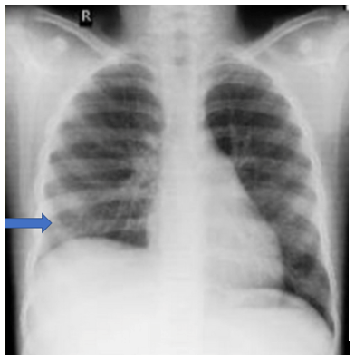
Diagnostic Assessment: Her hematological parameters were within limits except thrombocytosis. Serum biochemistry was normal. Liver and kidney function tests were within limits. Urine analysis was also normal. Therapeutic thoracocentesis was done and around 750ml of pleural fluid was aspirated gradually over the course of 48 hours following which she improved symptomatically. Pleural fluid analysis revealed a blood stained, sterile fluid, with protein content of 4.4 gm/dL, glucose of 91mg/dl, and LDH of 1043 IU/L. Fluid cytology revealed markedly increased lymphoid cell with plenty of RBCs. No malignant cells were visualized. Chest radiograph was suggestive of a massive right sided pleural effusion (Fig 1).
Figure 1: Chest x-ray showing right sided massive pleural effusion with mediastinal shift towards left.
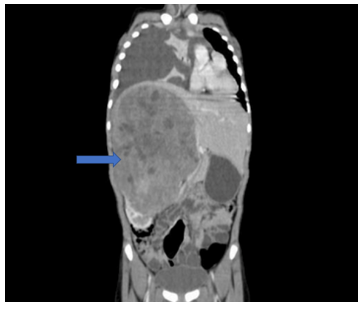
Contrast Enhanced Computed Tomography (CECT) of abdomen showed a large, heterogeneously enhancing mass (22x16x14cm) with multiple necrotic areas arising from the mid and upper pole of the right kidney (Fig 2).
Figure 2: CECT abdomen showing a large heterogeneously enhancing mass with necrotic areas in the right retroperitoneum having “claw sign” arising from the right kidney and extending across the midline to the left side with a normal left kidney. Right sided massive pleural effusion with adjacent passive atelectasis.
Diagnosis: On the basis of the above findings, a diagnosis of Wilms tumor with right sided pleural effusion was made.
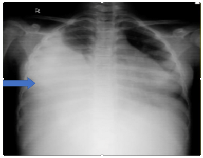
Therapeutic Intervention: As per the International Society of Pediatric Oncology (SIOP), her management plan included preoperative chemotherapy followed by radical nephrectomy and post-operative chemotherapy. She received 6 cycles of chemotherapy prior to surgery comprising of vincristine (1.5mg/m2), actinomycin D (45mcg/Kg) and adriamycin (50mg/m2). Her pleural effusion completely resolved after 4 weeks of chemotherapy without need of further thoracocentesis (Fig 3). She then underwent right radical nephrectomy. However, during surgery the mass was found to be densely adherent to the inferior venacava (IVC) throughout its length as well as to posterior aspect of liver and diaphragm. Some residual mass adherent to IVC was left behind. Histopathological examination of the specimen was suggestive of Wilms tumor (SIOP stage III) with no lymph nodal metastasis. In view of the residual disease, she received post-op radiotherapy with a total dose of 10.8 grays to the abdomen.
Figure 3: Chest x-ray showing complete resolution of pleural effusion after 4 weeks.
Follow up and outcome of the intervention: Further as planned, she was started on weekly chemotherapy with vincristine, actinomycin D and Adriamycin for 24 cycles. Currently, she has completed all her cycles with no further complications. Further plan of management includes surveillance ultrasonography for abdominal recurrence or development of a second primary tumor in the contralateral kidney and chest CT for pulmonary metastasis after three months.
Informed Consent- Informed written consent was obtained from her parents for publication of this case.
Discussion
The current case is interesting because of the unusual clinical presentation of pleural effusion. Since pleural effusion is the involvement of an organ system distant from the primary tumor site, there is a tendency to think of metastatic disease in such cases. However, there were no signs of primary pulmonary metastasis in this case. Therefore, the present case highlights that, those patients suffering from Wilms tumor presenting with pleural effusion need not be synonymous with metastatic disease and can have a favorable prognosis.
The most frequent site for metastasis in Wilms tumor is the lung, occurring in up to >90% of patients with metastatic disease. Rarely, pleural metastasis has also been documented. Pleural effusion is a rare presenting feature in children with Wilms tumor. The incidence of pleural effusion has been reported to be 4.3% [4]. Different mechanisms implicated in the causation of pleural effusion are – pleural metastasis, hypoproteinemia secondary to either chemotherapy or radiation induced transient liver injury or unrelated causes such as chylous exudate due to post-surgical lymphatic damage with associated infection [5]. Sympathetic effusion due to proximity of tumor to diaphragm or damage to it due to adhesion may be the cause in our case. Even though pleural effusion is seen in patients with Wilms tumor, massive effusions are rarely seen so as to cause respiratory distress as in our case. We were able to find at least 6 publications (17 children) of Wilms tumor with pleural effusion with pulmonary metastasis being reported in 4 children which are summarized in Table 1 [4-9]. The significance of pleural effusion in these groups of patients is the fact that it dramatically upgrades the staging of tumor and hence, changes the management of the patient. In a study by Wong JW et al [10], the malignant positivity of pleural effusion in WT with pleural effusion was found to be 35%. In stark contrast, a retrospective analysis done at St. Jude Children’s Research Hospital over 16-year period detected that there were no signs of metastasis in children with Wilms tumor presenting with pleural effusion [4]. The treatment modality of Wilms tumor with pulmonary metastasis includes chemotherapy with Vincristine, Actinomycin D and Adriamycin along with lung radiation therapy. The inappropriate upstaging of Wilms tumor leads to over-treatment with consequent treatment related toxicities. Pulmonary fibrosis and diffuse interstitial pneumonitis are complications secondary to lung radiation therapy for metastatic Wilms tumor. Dilated cardiomyopathy is a potentially life-threatening complication due to Adriamycin by virtue of its property of causing myocardial injury as well as it may act as a radiosensitizer which further potentiates the myocardial damage leading to reduced overall survival. Hence, appropriate staging as well as management are of utmost importance.
There is no consensus on the treatment of pleural effusion in Wilms tumor. Canopolat C et al have documented efficacy of chemotherapy alone in considerably resolving pleural effusion and noting a decrease in tumor size as well. Radiation therapy has also been documented to resolve pleural effusion [5]. In our patient, although a therapeutic thoracocentesis was performed to reduce the acute symptoms, the pleural effusion resolved completely by chemotherapy alone. Moreover, Computed Tomography thorax and pleural fluid cytology did not show evidence of any metastatic disease, hence, radiation therapy to lungs was not implemented.
Table 1: Summary of studies reporting pleural effusion in Wilms tumor.
| S.No |
Author (Year of publication) |
Patient demographics (Age in years; sex) |
Pleural fluid analysis |
Implicated etiology |
Treatment given |
|
1 |
Presented case |
11 years; Female |
Non-malignant effusion |
Unknown |
Therapeutic thoracocentesis followed by chemotherapy |
|
2 |
Corey B et al(2004) |
10 children |
Non – malignant effusion in all cases |
Unknown |
Chemotherapy alone in 8 cases; Additional pulmonary irradiation in 2 cases (Stage IV) |
|
3 |
Betkerur U et al (1977) |
Case 1: 4 year 8 months; male |
Malignant effusion |
Secondary to pleural metastasis |
Chemotherapy and radiation therapy |
|
Case 2: 5 year 4 months: female |
Chylous exudate, non-malignant |
Secondary to respiratory infection and damaged abdominal lymphatics |
Antibiotics alone with chemotherapy |
||
|
Case 3: 5 years; female |
Bilateral serous, non-malignant |
hypoproteinemia |
Spontaneous |
||
|
4 |
Kupeli S et al(2007) |
10 years; Female |
Non-malignant effusion |
Secondary to tru-cut biopsy |
Spontaneous resolution (although chemotherapy was being given) |
|
5 |
Al-Hadidi A et al |
12 years; Female |
Malignant effusion |
Secondary to pleural metastasis |
Chemotherapy and lung radiation therapy |
|
6 |
Canpolat C et al |
12 years; Female |
Malignant effusion |
Metastasis |
Chemotherapy alone |
|
7 |
Schinstine M et al(2006) |
9 years; Male |
Malignant effusion |
Secondary to metastasis |
Chemotherapy |
Conclusion
Although pleural effusion is a rare occurrence in cases of Wilms tumor, it need not be synonymous with metastatic disease and can be treated effectively with a good outcome. We recommend a careful strategy in cases presenting with pleural effusion so as to avoid chemotherapy and radiation therapy related morbidities. The lack of consensus on management of these groups of patients necessitates further studies in determining risk factors as well as management strategies.
Conflicting interests:The Author(s) declare(s) that there is no conflict of interest.
Funding: Not applicable
Informed consent: Written informed consent was obtained from the patient(s) for their anonymized information to be published in this article.
Ethical approval: Not applicable
Contributor ship: PZJ, ARR was a major contributor for writing this manuscript and patient care. AD was majorly involved in chemotherapy management. AA and KV were overlooking the patient’s management and corrected the final manuscript. JV critically reviewed the abstract section as well as the final manuscript. All the authors have read and approved of the final manuscript.
Acknowledgements: None
Abbreviations:
WT- Wilms tumor
vWF- von Willebrand factor
CT- Computed tomography
CECT- Contrast enhanced computed tomography.
SIOP- International Society of Pediatric Oncology
IVC - Inferior vena cava
List of Tables and Figures
References
- Pastore G, Znaor A, Spreafico F, Graf N, Pritchard-Jones K, et al. (2006) Malignant renal tumours incidence and survival in European children (1978–1997): report from the Automated Childhood Cancer Information System project. European journal of cancer 42(13):2103-14.
- Doganis D, Zborovskaya A, Trojanowski M, Zagar T, Bouka P, et al. (2019). Wilms tumour event-free and overall survival in Southern and Eastern Europe: Pooled analyses of clinical data from four childhood cancer registries (1999–2017). European Journal of Cancer 115: 37-46.
- Friedman AD (2013) Wilms tumor. Pediatr Rev 34: 328-330.
- Corey B, Yang CH, Wilimas JA, Davidoff A, Dome JS et al. (2004) Significance of pleural effusion at diagnosis of Wilms tumor. Pediatric blood & cancer 42: 145-148.
- Betkerur U, Lanzkowsky P (1977) Pleural effusion in Wilms' tumor. Journal of Pediatric Surgery 12: 523-525.
- Küpeli S, Varan A, Akyüz C, Ak?nc? D, Büyükpamukçu M, et al. (2007) Pleural effusion in wilms tumor after tru-cut biopsy. Pediatric Hematology and Oncology 24:555-558.
- Al-Hadidi A, Lapkus M, Novotny NM, Gowans LK, Chen PY, et al. (2020) Wilms Tumor with Pleural Metastasis. Global Pediatric Health 7: 2333794X20952292.
- Canpolat C, Jaffe N (1995) Wilms' tumor: cure of malignant pleural effusion exclusively with chemotherapy. Medical and pediatric oncology 24:274-277.
- Schinstine M, Abati A, Tsokos M, Fox E, Filie AC, et al. (2006) Cytological identification of metastatic epithelial nephroblastoma in pleural fluid: report of a case and review of literature. Diagnostic Cytopathology 34: 621-625.
- Wong JW, Pitlik D, Abdul-Karim FW (1997) Cytology of pleural, peritoneal and pericardial fluids in children. A 40-year summary. Acta cytologica 41: 467-473.