Anti-Inflammatory Perspective in the Pathogenetic Treatment of Malignant Brain Gliomas
Gridina N. Ya 1*
1 State Institution, “Institute of Neurosurgery named A.P. Romodanov of NAMS of Ukraine”, Kyiv, Ukraine.
*Corresponding Author:Gridina N. Ya, State Institution “Institute of Neurosurgery named A.P. Romodanov of NAMS of Ukraine”, Kyiv, Ukraine., Tel: +380 44 484 1873; Fax: +380 44 484 1873; E-mail:gridinanina@ukr.net
Citation: Gridina N. Ya (2022) Anti-Inflammatory Perspective in the Pathogenetic Treatment of Malignant Brain Gliomas. Cancer Prog Diagn 6: 129.
Received: September 04, 2022; Accepted: September 12, 2022; Published: September 15, 2022.
Copyright: © 2022 Gridina N. Ya. This is an open-access article distributed under the terms of the Creative Commons Attribution License, which permits unrestricted use, distribution, and reproduction in any medium, provided the original author and source are credited.
Abstract
Taking into account the role of inflammation in the pathogenesis of malignant gliomas, an approach has been developed to suppress the aggregation of blood cells in the second stage of the inflammatory process using low concentrations of verapamil hydrochloride. It has been shown that the progression of gliomas is accompanied by an increase in the aggregation of blood cells. For the first time, low concentrations of a 0.25% solution of verapamil hydrochloride (Farmak) were found to lower the level of blood cell aggregation and is not a toxic drug. An experiment on rats showed that inoculation of grafted 101.8 glioma cells (an analogue of human glioblastoma) into the brain of fetal rats on the 14th day of pregnancy leads to complete death of glioma cells after 7 days, which was confirmed by morphological studies. The use of verapamil hydrochloride in patients with glioblastoma contributed to a significant increase in life expectancy compared with the group of patients who did not receive treatment with low concentrations of verapamil hydrochloride.
Key Words:
Malignant Gliomas; Tumor-Associated Inflammation; Surface Plasmon Resonance; Verapamil Hydrochloride; Low Concentrations; Grafted Glioma of Rats 101.8, Grafting of Glioma Cells 101. 14-Day-Old Fetuses of Rats
Introduction
The problem of treating malignant brain tumors is very complex in oncological practice, comparable to the treatment of melanoma, pancreatic cancer, seminoma, and other most malignant types of neoplasms. This condition is largely due to the fact that the main types of chemotherapy and other treatment approaches are not epipathogenetic, i.e., not always take into account the mechanisms of tumor growth. To develop a treatment for these types of tumors, it is necessary to understand the mechanisms of their pathogenesis not only at the molecular, but also at the systemic levels.
One of the misconceptions about the pathogenesis of malignant gliomas is the belief that malignant tumors are a typical pathological process. However, malignant tumors progress in close association with an inflammatory process called tumor-associated inflammation (TAI). For a long time, the presence of a significant role of TAI was not given much importance. The fact is that generally accepted laboratory methods for determining inflammation in the body are insensitive and do not always determine the presence of micro-inflammation, which in essence is TAI. Only the use of modern high technologies makes it possible to determine the presence of inflammation in the body, expressed in the form of micro-inflammation.
Cases of occurrence of gliomas after gunshot wounds and mechanical non-penetrating traumas of the skull are described [1]. In 24% of patients with cerebral gliomas, a history of traumatic brain injury was noted. On average, brain tumors occurred 3 months to 12 years after TBI, and in some cases, tumors occurred even 15–20 years after injury [2–3]. There are no serious concepts explaining these observations.
The second misconception was the idea of tumor growth from the cells of parenchymal organs. With the discovery of stem cells, the mechanism of tumor growth, which was not as simple as previously thought, became clear, associating it with the multiplication of microorganisms. Indeed, the tumor grows from cells, but from what? What mechanism causes them to multiply in this particular organ, ignoring the signals of the prohibition of independent reproduction? We have just begun to understand the phenomenon of these processes, perhaps not completely and not always correctly.
The author proposes to consider the growth of malignant tumors, including gliomas, as a pathological regenerative process carried out under conditions of constant action of microinflammation on mesenchymal stem cells of the bone marrow and, possibly, stem cells of the organ parenchyma. Mesenchymal cells migrating to a lesion in a parenchymal organ may initially be pathologically altered; in addition, they continue to be further destroyed in the lesion itself, since the mechanism of their protection in the form of an epithelial-mesenchymal transition does not work [4]. Therefore, instead of regenerating the destroyed tissue of the parenchymal organ to normal cytoarchitectonics, mesenchymal cells can form a tumor conglomerate. Moreover, in most types of tumors, except intracerebral, stem cells migrate inside the body, settling in other damaged foci, mimicking the picture of metastasis.
With prolonged exposure of microinflammation to stem cells, they can change from multipotent and pluripotent to totipotent. Hence the markers of "pregnancy", which baffle many oncologists. Therefore, having at least an approximate understanding of the mechanism of tumor growth and progression of tumors, it becomes clear that blocking the inflammatory process in the body, correcting the hormonal status, creating conditions for normalizing the processes of repair and regeneration will lead to long-term remission in malignant tumors or to a complete cure of the body. Attention should be paid to the fact that inflammation and early embryogenesis are processes that occur at different time intervals. In the first half of embryogenesis, when inflammatory reactions are not observed due to the absence of a formed vascular system, malignant tumors do not arise. In the fetuses of the second half of pregnancy, newborns and young children, tumors are usually benign and respond well to combined treatment. The maximum of tumors occurs in the adult population, especially in the elderly, when many ischemic foci appear in the tissues, but since the functions of the inflammatory process are already fading, malignant tumors become sluggish.
The aim of the work was to demonstrate the possibilities of a new technology of surface plasmon resonance in determining inflammation in brain gliomas and the effect of verapamil hydrochloride at low concentrations on the life expectancy of patients with glioblastomas, as well as to study in an experiment on live fetuses of rats (14 days of pregnancy) the possibility of survival of rat cells transplanted glioma 101.8 in the absence of manifestations of the inflammatory process.
Materials and Methods
The generally accepted clinical methods of blood tests (ESR, C - reactive protein, the number of cellular elements in the blood) showed the absence of an inflammatory process in patients with gliomas of II - IV grades of malignancy. Determination of indicators of aggregation of blood cells on the biosensor "Plasmon". Indicators of blood cell aggregation (SPR - indicators) were determined in 52 healthy individuals and 239 patients with gliomas of various grades of malignancy (Table 1) before their treatment in a neurosurgical clinic.
Table 1: Number of examined healthy individuals and patients with neurosurgical pathology.
|
Diagnosis |
Number of People examined |
|
Healthy |
52 |
|
Glioma of ?? grade of malignancy |
52 |
|
Glioma of ??? grade of malignancy |
103 |
|
Glioma of IV grade of malignancy |
84 |
The effect of surface plasmon resonance (SPR) [5–7] uses a new direction in biosensorics, which made it possible to study the initial processes of blood cell aggregation in real time. The device allows you to register intercellular interaction at nanoscale distances (about 200-300 nm).
The principle of operation of the device is that a collimated polarized beam of an optical excitation source, falling from the side of the glass onto the chromium deposited through the intermediate adhesive layer (1-1.5 nm), onto the gold nanoscale layer (45-50 nm) can, at certain angles of incidence, excite plasmon oscillations in the gold layer. The fact and parameters of excitation of plasmon oscillations can be fixed by determining the parameters of the light reflected from the gold film. The conditions for the excitation of plasmon oscillations essentially depend on the medium in contact with the gold film. Any changes in this medium lead to a change in the angle of incidence of light, at which plasmon oscillations are excited. In our studies, in particular, blood cells were in contact with the surface of the gold film. The developed software makes it possible to use the capabilities of the devices to the maximum when working in laboratory conditions.
The plasmon resonance method has a significant advantage over all other methods for determining the degree of aggregation of blood cells. This advantage lies in the real opportunity to conduct studies on native blood cells without the addition of sodium chloride solutions or various buffer systems, as required by the conditions for determining cell aggregation in other methods. The discovery of ion channels and ionotropic receptors on blood cells has been made in the last decade. Many methods for determining the aggregation or zeta potential of blood cell membranes had already been developed and widely used by that time. The researchers did not even suspect that buffer solutions containing various electrolyte salts affect the transmembrane potential and blood cell aggregation, block various channels and change the activity of ionotropic receptors. Therefore, the results of studies to determine the degree of aggregation of blood cells using saline solutions or buffer systems may not correspond to reality.
The SPR indicator is considered to be the shift of the resonance curve of the SPR, measured in degrees.
To study the effect of low concentrations of verapamil hydrochloride on the level of blood cell aggregation (SPR indicators), a 0.25% solution of verapamil hydrochloride was dissolved from 10 to 10,000 times with ion-free water. To 200 μl of peripheral blood placed in microtubes, 20 μl of verapamil hydrochloride was added, each separately from the obtained dilutions. Then these samples were examined on the device to determine the parameters of the SPR. We analyzed at what dilution of verapamil hydrochloride the aggregation of blood cells decreased to the maximum.
Animal experiments with grafted glioma 101.8
In the first series of experiments, 1 million cells of transplanted glioma of strain 101.8 were inoculated into white rats of 3-4 weeks of age (50 rats) of the Wistar line.
In the second series of experiments, the tumor was transplanted into the brain of 14-day-old fetuses of rats according to the method developed by the author. In pregnant rats (15 rats) under hexenal anesthesia, the abdominal cavity was opened, the horns and body of the uterus were removed, and they were placed on a sterile napkin moistened with warm saline. Tumor cell suspension (about 105 cells) was injected into the left parietal region of each fetus with a micro syringe. Then the uterus was placed in the abdominal cavity, the abdominal wall was sutured in layers. Six days after tumor inoculation, the brain was removed from viable fetuses, fixed in 10% neutral formalin, and embedded in paraffin-celloidin. Serial sections of the brain of embryos were stained with hematoxylin-eosin and picrofuchsin and studied under a microscope.
Determination of the life expectancy of patients with malignant gliomas in the treatment of low concentrations of verapamil hydrochloride
Patients with malignant gliomas of the brain in the postoperative period after courses of combined treatment were prescribed treatment with low concentrations (10,000-fold dilution) of verapamil hydrochloride. Patients took the drug diluted with water three times a day 20 minutes before meals for life, without interruption. The life expectancy of patients treated with verapamil hydrochloride and not taking the drug was studied.
Statistical processing of the obtained data was carried out in the Statistica-5.5 package using nonparametric data evaluation methods. The correctness of the distribution of signs for each of the obtained variational series, the average values for each sign that were studied, and standard deviations were evaluated. The significance of the difference in values between independent quantitative values using the Man-Whitney U-test, and between dependent ones using the Wilcoxon test.
Results and Discussion
In the study of the level of aggregation of blood cells using a highly sensitive sensor device "Plasmon", statistically significant differences in the parameters of PPR were obtained in patients with brain gliomas of various degrees of malignancy and in patients after moderate craniocerebral injury. There is a regular decrease in the charge of blood cell membranes in patients compared with a group of healthy people. The assumption is confirmed that the growth and progression of malignant gliomas is accompanied by a decrease in the transmembrane potential, mediated by the level of blood cell aggregation, from a more benign glioma phenotype (grade II) to a highly malignant phenotype (grade IV).
Figure 1: Characteristics of blood cell aggregation (i.e., values of the angular shift of the SPR peak position) typical for patients with various degrees of malignancy.
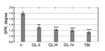
Note: ** ? ≤ 0.01; *** ? ≤ 0.001.
K – blood samples without additions of preparations; GL. II, GL.III and GL. IV – blood samples for the cases of gliomas of the II, III, IV grades of malignancy, respectively; TBI – the case of trauma brain injury. In traumatic brain injury, a decrease in transmembrane potential is a short-term phenomenon, but there are distant consequences in the form of growth of tumors in the brain [2-3]. A study of the effect of verapamil hydrochloride in various dilutions showed the following results.
In the group of patients with gliomas of IV grade of malignancy, when diluted to 10-1 degrees, promotes an increase in the degree of aggregation of blood cells (Figure 2).
Figure 2: Effect of different dilution of verapamil-anti-inflammatory drug (from 10-1 to 10-6) on the transmembrane potential (TMP) level by SPR blood cells aggregation method in glioma malignancy patients.
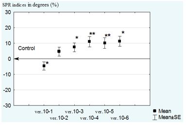
At dilutions of 10-3, 10-4, 10-5 and 10-6 degrees, the drug significantly reduces blood cell aggregation compared to the control: 10-4, 10-5 degrees - with reliability (p≤0.01).
Therefore, high-dose dilutions of verapamil contribute to an increase in blood cell aggregation in neurosurgical pathology, and low-dose concentrations of the drug, on the contrary, lower the level of blood cell aggregation and, therefore, reduce the manifestations of the inflammatory process in the body, since blood cell aggregation is stage II of inflammation.
When comparing the SPR parameters in gliomas of various grades of malignancy, it should be noted that with an increase in the degree of malignancy, the effectiveness of verapamil in relation to the reduction of blood cell aggregation increases. With glioma of IV grade of malignancy the highest rates of SPR are noted, therefore, there is a maximum decrease in blood cell aggregation.
Experimental studies on rats
In the second series of experiments, 101.8 glioma cells were inoculated directly into the brain tissue of 14-day-old fetuses of rats with the condition that the further course of rat pregnancy continued. The day before birth, the fetuses were removed from the uterus and the brain tissue was studied by morphological methods. The experiments carried out made it possible to observe the result of direct contacts between tumor and brain cells of fetal rats in vivo. Histological examination of brain sections of fetal rats revealed small clusters of 101.8 glioma cells with destroyed cytoplasm.
Nuclei of many cells were hyper- or hypochromic, with signs of pycnosis and karyolysis. At the same time, the tissue of the embryonic brain in the circumference of the puncture passage and throughout the rest of its length retained its usual structure and architectonics, similar to the control. As a result of the experimental studies, it should be concluded that the absence of an inflammatory process in the body of fetal rats by the 14th day of embryogenesis leads to suppression of the growth of glioma 101.8 as a result of the destruction of tumor cells in the environment of the fetal tissues. At the same time, the inoculation of 101.8 glioma cells in mature brain tissue leads to the death of experimental animals as a result of the presence of an inflammatory process in the trans-barrier organ, the brain.
Therefore, the suppression of the inflammatory process, most often TAI, by any available means will contribute to the inhibition of the growth of malignant gliomas.
Determination of life expectancy in patients with malignant gliomas treated with low concentrations of verapamil hydrochloride
The control group, which received only traditional combined methods of treatment of malignant gliomas, included 39 patients. In the group of patients, after traditional courses of chemotherapy and radiation, receiving low concentrations of verapamil hydrochloride, 13 patients were included.
Table 2: Life expectancy of patients with glioblastomas in the postoperative period with using of verapamil- hydrochloride in low concentrations in order to inhibit the inflammation process.
|
Group |
Number |
Mean |
Standard error of mean |
Median life expected |
|
Control |
39 |
9,30 |
1,04 |
8,00 |
|
Verapamil |
13 |
22,46 |
2,23 |
19,00 |
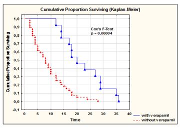
The systematic suppression of the TAI in patients with glioblastomas in the late postoperative period led to an increase in their life expectancy [9-11]. These studies showed results in which the average life expectancy increased up to 22 months after surgery, compared with the average (9 months) in the group in which verapamil-hydrochloride was not taken (Table 2) (Figure 3). Three patients did not take courses of chemotherapy and radiation. Their life indicators were significantly higher than in the group of patients after combined treatment of glioblastomas.
Figure 3: Differences between groups of patients who received and did not receive verapamil treatment are significant by the F - Cox test. The significance level is 0.001.
Summary
The paper presents data indicating a significant role of chronic microinflammation in the pathogenesis of malignant brain gliomas. Unfortunately, the issues of pathological changes in stem cells under conditions of chronic inflammation are poorly covered in the literature [12]. Therefore, the mechanisms of these processes remain to be explored. It can be concluded that one of the main mechanisms of malignant progression of gliomas is pathologically altered regeneration of tissue and organ cells as a result of its implementation under conditions of chronic microinflammation. This conclusion is supported by the presented studies. Unfortunately, it is not possible to completely slow down inflammation in the body, because microinflammation is observed even in healthy people, which has physiological characteristics. Therefore, the approach to the treatment of malignant gliomas should also affect other systems in the body associated with the correction of the relationship between repair, carried out by the mechanisms of inflammatory genesis, and regeneration mechanisms, which are carried out mainly by stem cells and about which our knowledge is still far from perfect. I would like to believe that the presented results of the studies will convince oncologists to carry out anti-inflammatory measures using a calcium channel blocker in low concentrations of verapamil, as a drug that does not have side effects with long-term use, unlike hormonal drugs, etc.
References
- Salvati M, Caroli E, Rocchi G, Frati A, Brogna Ch, et al. (2004) post-traumatic glioma. Report of four cases and review of the literature. Tumori 90: 416 - 419. [Crossref]
- Dvorak HF (1986) Tumors: wounds that do not heal. Similarities between tumor stroma generation and wound healing. New Engl. J.Med 315: 1650 - 1659. [Crossref]
- Kudravi SA, Reed MJ (2000) Aging, cancer, and wound healing. In vivo14: 83 - 92. [Crossref]
- Thiery JP (2002) “Epithelial-mesenchymal transitions in tumour progression”. Nat Rev Cancer 2: 442-454. [Crossref]
- Bondeson K, Frostell-Karlsson A, Fagerstam L, Magnusson G (1993) Lactose Repressor-Operator DNA Interactions: Kinetic Analysis by a Surface Plasmon Resonance Biosensor. Analyt. Biochem 214: 245 - 251. [Crossref]
- Gridina N, Dorozinsky G, Khristosenko R, Maslov V, Samoylov A, et al. (2013) Surface Plasmon Resonance Biosensor. Sensors&Transducers 149: 60-68.
- Dorozhinsky GV, Maslov VP, Ushenin YuV (2016) Sensor devices based on surface plasmon resonance. Monograph. NTUU "KPI" Kyiv 264
- Clements JD, Lester RAJ, Tong G, Jahr CE, Westbrook GL, et al. (1992) The time course of glutamate in the synaptic cleft. Science 258: 1498- 1501.
- Gridina N Ja, Biloshitsky VV, Morozov AN, Rozumenko VD, Draguntzova NG, et al. (2014) The new approaches to use verapamil and ketamin at brain glioma therapy. Ukr Neurosurgery journal 4: 17-21.
- Gridina NYa, Shvachko LP, Draguntsova NG (2016) “Tumor-Associated Inflammation Mechanisms Correction by Verapamil at Brain Gliomas Progression”. Eur J Pharmaceutic Med Res (EJPMR) 3: 73-78.
- Gridina NYa, Morozov AN, Rozumenko VD, Uschenin YuV, Shvachko LP, et al. (2020) “Some aspects of the Systemic Mechanism of Brain Malignant Gliomas Progression and Methodological Approaches to its Correction”. EC Neurology 12: 80-90. [Crossref]
- Tanno T, Matsui W (2011) “Development and maintenance of cancer stem cells under chronic inflammation”. Journal of Nippon Medical School 78: 138-145. [Crossref]