Xeroderma Pigmantosum with Multiple Malignant Lesions, Present in Siblings Linking it to Genetic Aspect of the Disease, and Possible Treatments
Dr Dipen Shivanuja 1*
1 Gujarat cancer and Research Institute, B. J.Medical college, Ahmedabad, Gujarat, India
*Corresponding Author:Dr.Dipen Shivanuja, Gujarat cancer and Research Institute, B. J.Medical college, Ahmedabad, Gujarat, India, Tel: +91- 9518438131; Fax: +91- 9518438131; E-mail:dipenmistry57555@gmail.com
Citation: Dipen Shivanuja (2022) Xeroderma Pigmantosum with Multiple Malignant Lesions, Present in Siblings Linking it to Genetic Aspect of the Disease, and Possible Treatments. Cancer Prog Diagn 6: 127.
Received: July 28, 2022; Accepted: August 20, 2022; Published: August 24, 2022.
Copyright: © 2022 Dr Dipen Shivanuja. This is an open-access article distributed under the terms of the Creative Commons Attribution License, which permits unrestricted use, distribution, and reproduction in any medium, provided the original author and source are credited.
Abstract
Being a Geno dermatosis, photosensitive disorder XERODERMA PIGMANTOSUM (XP) is a rare autosomal recessive disorder. Patient with XP commonly presented with an interactable and dermatological disease. Patients with XP are prone to cutaneous, mucosal malignancies and ophthalmic complications. Reason being inability to repair the ULTRAVIOLET (UV) radiation dispensed damage to Deoxyribonucleic Acid (DNA), genetic nature of disease is revealed. Not only genetic but environmental factor plays big role in course of disease. In XP cutaneous and mucosal basal cell carcinoma (BCC) And squamous cell carcinoma (SCC) or melanoma is most common.
XP characterized with sever sunburns, development of many freckles at early age, rough surfaced wart like growths, cutaneous and mucosal malignancies, blistering, telangiectasis, ophthalmic manifestations, limited growth of hair on chest and legs and dry skin. 45 to 60% patients of XP were observed to be developing malignant skin neoplasm in different studies [1]. compare to normal controls Robbins has shown that incident of internal neoplasm in XP patient was 10-20 times higher. Angiosarcoma and Fibrosarcoma have also been reported. In XP eye tissues are also exposed to UV and cause lesion mainly in anterior ocular segment, such as conjunctival xerosis, corneal drying, decrease of lacrimation, corneal neovascularization, ectropion, blepharitis, pigmentation of conjunctiva, cataract etc. As most of UVB could not reach to retina chances of morbid changes are very less but abnormalities in optic nerve as neurological symptoms can manifest.[2]
Introduction
The disease represented by evident photosensitivity, premature onset of all major type of skin cancers was First described by HEBRA AND KAPOSI in 1874.[3] It was reported for the first time that XP is genetic in nature in which genetic abnormalities occur at DNA level. Nucleotide Excision Repair enzyme is mutated, and human cells are unable to get repaired because of lack or reduction of NER enzyme. NER enzyme repair damages happened by UV at DNA level. XP occurs worldwide including all countries, all races and all ethnic background. Worldwide incidence of XP is 1 in 1000000, in united states of America (USA) and Europe it is 1 in 250000[4] whereas in Japanese people its six-fold more. [5] The exact incidence of XP in India is not known but it is reported that occurrence is more in south Indian regions, especially Karnataka.[6] XP typically diagnosed at early age but with advanced technology we can diagnosed it prenatally with help of amniocentesis studies and chorionic villous sampling. We also can measure DNA repair factor in blood or from skin.
We present a case of siblings, younger brother a 9-year-old male child and an elder sister a-13-year-old female child with xeroderma pigmentosum. Both siblings have cutaneous and mucosal malignant lesions.
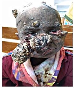
Younger brother a-9-year-old male child presented initially with swellings on lip, scalp, face and multiple hyperpigmented skin lesions over whole body as a diagnosed case of xeroderma pigmentosa. Further investigation revealed squamous cell carcinoma right side of cheek, pyogenic granuloma in lip lesion and no evidence of malignancy from scalp lesion then further taken chemotherapy For response computed tomography done which shows lesions on skin of cheek on both side and nose with soft tissue thickening on tip of tongue but pathologically no evidence of malignancy on tip of tongue. Then taken multiple chemotherapy regimen. For response assessment computed tomography was done which shows increase in size of previous lesion. Another regimen of chemotherapy taken as further line of treatment. Further workup done computed tomography shows additional lesions over the bilateral parietal region of scalp. For further management he was referred for radiotherapy. Radiotherapy was given for scalp lesion.
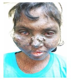
Elder sister a 13-year female child presented with a lesion over tongue, lower lip and as described above multiple hyperpigmented skin lesions as diagnosed case of xeroderma pigmentosa. Punch biopsy from tongue and lip shows well differentiated squamous cell carcinoma. As further investigated computed tomography and magnetic resonant imagine was done which shows the lesion on the left lateral border of tongue then taken three cycles of chemotherapy. Lesion was clinically completely responded. After 1 month she developed another new lesion on scalp. Computed tomography brain was done which shows lesion on right parietal occipital lesion. wide local excision was done which shows benign lesion suggestive of telangiectasis. Referred for radiotherapy for primary lesion management. Curative radiotherapy was given for tongue lesion. after 2 month she developed ulcer over left side cheek. Biopsy was taken which shows squamous cell carcinoma. After that she developed multiple swelling over face, forehead and scalp. Biopsy was done from left inner canthus shows basal cell carcinoma so further investigated. Computed tomography and magnetic resonant imagine was done which shows lesion involving skin and subcutaneous plane of right premolar region. So as further treatment 3 cycles of chemotherapy were given.
Case 1 Case 2
|
case |
Genetic history |
Malignancy |
Genetic disorder |
Prognosis |
Treatment |
|
Case 1 |
present |
Squamous cell carcinoma tongue |
Xeroderma pigmentosa |
Poor |
Chemotherapy Radiotherapy |
|
Case 2 |
present |
Squamous cell carcinoma scalp, cheek |
Xeroderma pigmentosa |
Poor |
Chemotherapy Radiotherapy |
Discussion
In XP there are 7 gene type has been recognized which are given name alphabetically. (XP-A XP-B XP-C XP-D XP-E XP-F XP-G).[7] Here XP-C and XP-E recognizes the photoproduct in DNA. XP-B and XP-D are part pf protein complex TFIIH which opens up the structure of DNA around the side of photoproduct. XP-A protein verifies that proteins are in correct position and then the nucleases XP-G and XP-F cut the DNA on either side of damage, so damage is removed and replaced with healthy proteins. Patient with defect in XP-C and XP-E gene does not in general shows extremes of abnormality.
XP happened to be inherited disease; a radical cure is cannot be expected. The premises of XP patient care are defense from UV and symptomatic treatments for complications. Early diagnosis, Strict avoidance of sunlight, minimize UV exposure by lifestyle changes such as wearing protecting clothing, glasses, using protective shields in windows, applying sunscreen lotions, using umbrellas when stepping out. Genetic counselling plays important role in cases of XP. Surgical excision is modality used for various cutaneous and mucosal lesions. Oral 13-cisretinoic acid reportedly reduces the risk of new cancers. Cryotherapy is also one of the treatment techniques.
Fluorouracil pyrimidine analogues are also used to treat certain types of cancer. Reports are presented that imiquimod is useful for actinic keratosis and basal cell carcinoma. Interferon α is useful for melanoma. [8,9]
Frequent follow up with doctors handling the case is encouraged every 3-6 months for sack of early detection of new lesions and to check ophthalmological manifestations.
For local relief Betadine ointments for wart type lesion to prevent infection and Neosporin power application is also being practiced. Less than 40% of the individual with the disease survive beyond age of 20, some less severe cases make it up to 40 years and so.
Conclusion
Xeroderma pigmentosa is a rare type of autosomal recessive genetic disorder. Patient with XP are prone to cutaneous, mucosal malignancy and ophthalmic manifestation. The main reason XP is inability to repair the UV radiation inflicted damage to DNA. In XP hypo and hyper pigmentation present with gradually increase in size in number. They are prone to sunburns and photosensitive damages. Small warty growths are seen over face and expose areas.
Declaration of Patient Consent
The authors certify that they have obtained all appropriate patient consent forms. In the form the patient(s) has/ have given his/her/their consent for his/her/their images and other clinical information to be reported in the journal. The patients understand that their names and initials will not be published, and due efforts will be made to conceal their identity, but anonymity cannot be guaranteed.
References
- Bandyopadhyay R, Nag D, Bandyopadhyay S, Sinha SK (2012) Atypical fi broxanthoma: An unusual skin neoplasm in xeroderma pigmentosum. Indian J Dermatol 57: 384-386. [Crossref]
- Shinichi Moriwaki,' Fumio Kanda, Masaharu Hayashi, Daisuke Yamashita, Yoshitada Sakai.' Chikako Nishigori, "Xeroderma pigmentosum clinical practice guidelines revision committee.
- Halpern J, Hopping B, Brostoff J (2008) Photosensitivity, corneal scarring and developmental delay: Xeroderma Pigmentosum in a tropical country. Cases journal 1: 254. [Crossref]
- Robbins JH, Kraemer KH, Lutzner MA, Festoff BW, Coon HG, et al. (1974) Xeroderma pigmentosum: an inherited disease with sun-sensitivity. multiple cutaneous neoplasms, and abnormal DNA repair. Annals Internal Med 80: 221-248. [Crossref]
- Hirai Y, Kodama Y, Moriwaki S, Noda A, Cullings HM, et al. (2006) Heterozygous individuals bearing a founder mutation in the XPA DNA repair gene comprise nearly 1% of the Japanese population. Mutat Res 601:171-178. [Crossref]
- Rita V Vora, Rahul Krishna Suresh Kumar Kota, Nilofar G Diwan, Shree Krishna VD (2016) Shree Krishna Hospital, Anand, Gujarat, India.
- Stefanini M. Kraemer (2008) KHK: Xeroderma pigmentosum. In Neurocutaneous Diseases Edited by: Ruggieri M. Pascual Castroviejo I. di Rocco C 51: 771-792.
- Yarosh D, Klein J, O'Connor A, Hawk J, Rafal E, et al. (2001) Effect of topically applied T4 endonuclease V in liposomes on skin cancer in xeroderma pigmentosum: a randomized study Xeroderma Pigmen tosum Study Group. Lancet 357: 926-929. [Crossref]
- Turner ML, Moshell AN, Corbett DW, Stern JB, Roth MJ, et al. (1994) Clearing of melanoma in situ with intralesional interferon alfa in a patient with xeroderma pigmentosum. Arch Dermatol 130: 1491-1494. [Crossref]