Melanocyte Growth Factor Deprivation is Intertwined with Oxidative Stress and Autoimmune Theories in The Etiology of Vitiligo. A Basis for Treating Vitiligo
Ramaiah Abburi*1
1Samastha Laboratories Hyderabad, India
*Corresponding Author:Ramaiah Abburi, Samastha Laboratories Hyderabad, India, TEL:+91-9012952001 ; FAX:+91-9012952001; E-mail:ramaiah_abburi@yahoo.com
Citation:Ramaiah Abburi (2017) Melanocyte Growth Factor Deprivation is Intertwined with Oxidative Stress and Autoimmune Theories in The Etiology of Vitiligo. A Basis for Treating Vitiligo. J Dermatol & Ther 2:107.
Copyright:© 2017 Ramaiah Abburi, et al. This is an open-access article distributed under the terms of the Creative Commons Attribution License, which permits unrestricted use, distribution, and reproduction in any medium, provided the original author and source are credited
Received date: November 18, 2017; Accepted date: December 14, 2017; Published date: December 17, 2017.
Double Blind
Double Blind Randomized Phase IV Clinical Trial on bFGFRP in Vitiligo
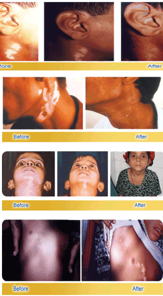
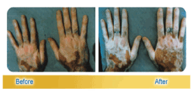
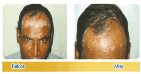
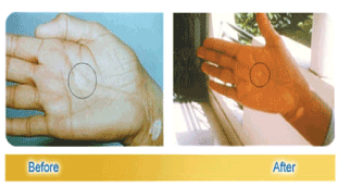
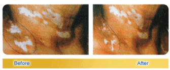
All the above described clinical trials on bFGFRP were open ended. In view of the fact that western countries require only double blind clinical trials on any prospective drug for it to be approved as a drug, a multi-center double blind clinical trial was done on bFGFRP in combination with NBUVB or without. Two macules in non-sun exposed areas were selected on each patient with stable non-segmental vitiligo. The duration of treatment of volunteers was for 3 months starting with or without the topical application of bFGFRP followed by NBUVB on 30 patients. The results demonstrated that bFGFRP acted synergistically with NBUVB in repigmentation of the macules. More repigmentation occurred with NBUVB + bFGFRP at all-time points of evaluation (1, 2, and 3 months) than with NBUVB alone. NBUVB+ bFGFRP was superior to NBUVB alone in repigmenting vitiligo macules (23). Thus, it is clearly demonstrated that bFGFRP can be successfully used by itself or with synergistic effect in combination with many other modalities of treatment to treat vitiligo.
Table-1: Growth Characteristics of Normal and Vitiligo Melanocytes.
| Normal | Uninvolved from Vitiligo | Perilesional from Vitiligo | Uninvolved of Repigmenting | Perilesional from Repigmenting | |
| Initial seeding capacity | 124#12.38 | 53.8#10.1 | 27.6#3.92 | 168.9#12.4 | 134#1.6 |
| Lag in growth (d) | 0 | 6-11 (d) | no growth | 0 | 0 |
| Doubling time | 8.1#1.2 | 8.5#.89 | No growth | 4.6#0.47 | 4.25#0.25 |
| Number of passages | Four | No passagebility | No growth | >9 | >9 |
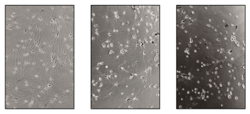
Figure 1: The Melanocytes from Normal, the perilesional &far away from the vitiligo macule.
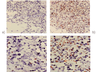
Figure 2: bFGF increased the proliferation of melanocytes in mixed cultures of keratinocytes and melanocytes from vitiligo patients.

Figure 3: Guinea pig ears before and after depigmentation.
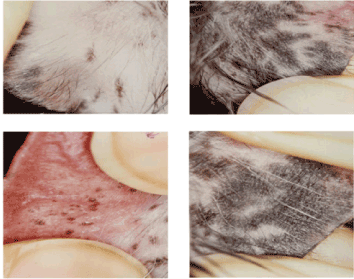
Figure 4: Effect of bFGFRP on the repigmentation of depigmented patches of guinea pig ear lobes compared to controls.
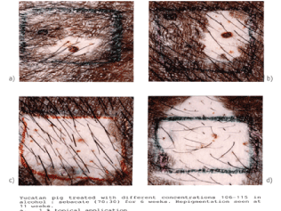
Figure 5: Topical application of (bFGFRP) at varying concentrations on depigmented patches of skin of Yucatan swine.

Major Conclusions
Growth factor deprivation theory as a possible cause of vitiligo (3) is in the light of the present literature mentioned above intimately intertwined with other prevailing theories of oxidative stress and auto immune theories (5). These theories are not mutually exclusive but interrelated and all together contribute to the loss of melanocytes resulting in vitiligo.
Acknowledgements
ACK The author is grateful to Prof. Davinder Prasad of PGI Chandigarh who supervised the clinical trials while he was at Himalayan Institute of Medical Sciences Jolly Grant with deca peptide lotion alone, PUVASOL with and without deca peptide lotion and with oral mini pulse therapy with and without deca peptide lotion and Prof AS Kumar at Owaisi hospital Hyderabad who supervised the clinical trial with PUV-A+deca peptide lotion on vitiligo patches resistant to PUV-A alone and above all to all the volunteers who participated in the various clinical trials described herein.