Schwanomma of The Left Iliohypogastric Nerve: A Case Report and Technical Consideration
Md. Shafiul Alam1*, Kaisar Haroon1, Tayseer Farzana2, Forhad Ahmed3, Taher Taher4
1 Associate Professor, Department of Neurosurgery, National Institute of Neurosciences & Hospital, Dhaka, Bangladesh.
2 Consultant, Department of Radiology & Imaging, Popular Diagnostic Center, Mirpur, Dhaka, Bangladesh.
3 Medical Officer, Department of Neurosurgery, National Institute of Neurosciences & Hospital, Dhaka, Bangladesh.
4 Assistant Professor, Holy Family Red Crescent Medical College Hospital.
*Corresponding Author:Md. Shafiul Alam, Associate Professor, Department of Gamma Knife, National Institute of Neurosciences & Hospital, Dhaka, Bangladesh, Tel: 01711567358; Fax: 01711567358; E-mail: dr_chapal@hotmail.com
Citation: Shafiul Alam MD, Kaisar Haroon, Tayseer Farzana, Forhad Ahmed, Taher Taher, et al. (2020) Schwanomma of The Left Iliohypogastric Nerve: A Case Report and Technical Consideration. Archiv Neurol Neurosurgery3: 121.
Copyright: © 2020 Md. Shafiul Alam, et al. This is an open-access article distributed under the terms of the Creative Commons Attribution License, which permits unrestricted use, distribution, and reproduction in any medium, provided the original author and source are credited.
Received: October 24, 2020; Accepted: November 06, 2020; Published: November 09, 2020.
Abstract
Schwannomas are encapsulated tumors arising from Schwann cells of the nerve sheath, and are usually solitary sporadic lesions. The schwannoma of the ilio-hypogastric nerve specially in the retro-petoneal space is very rare. We are reporting a case of the schwannoma of the left ilio-hypagastric nerve over the psoas muscle. The diagnosis was done by ultarsonogrsphy and MRI. The histopathological study confirmed the diagnosis. The tumour was excised through retro-peritoneal approach. By this approach total excision of the tumour with preservation of ilio-hypogastric nerve is possible.
Keywords
Ilio-hypogastric nerve, Schwanomma, retroperitoneal approach, resection.
Introduction
Schwannomas are encapsulated tumors arising from Schwann cells of the nerve sheath, and are usually solitary sporadic lesions [1]. Schwannomas are rarely found in the retroperitoneum, accounting for only 0.7% to 2.7% of all primary schwannomas [1]. They can be found in any nerve trunk, except for cranial nerves I and II, and their usual location is the head, neck, the flexor surfaces of the extremities and the posterior mediastinum or the retroperitoneum2. The first peripheral nerve tumor to satisfy the currently acceptable clinical and histological criteria of this tumor was described in 1910 by Verocay, who gave it the name of neurinoma. The more descriptive terms of neurilemoma or neurilemoma were introduced by Stout [3].
Schwannoma usually arises between the third and sixth decades of life, with an equal predilection for men and women4. Sachwannoma is a benign tumour. Malignancy in a solitary schwannoma is rare [5].
When tumours are slow-growing benign tumour with favourable prognosis, they are called ancient schwannoma. These lesions begin as solid tumours but as they become larger, they undergo spontaneous degeneration with areas of haemorrhage and cystic changes [6]. The tumor is classically described in two forms. The fascicular type (Antoni A) is composed of compact spindle-shaped cells that are arranged in short bundles or interlacing fascicles. There may be nuclear palisading and formation of Verocay bodies. Areas of fibrosis, necrosis, and hemorrhage may occur with thrombosis of the blood vessels. The reticular type (Antoni B) is highly vascularized and shows a loose arrangement of the Schwann cells in an open network of cysts and reticular fibers. There is no palisading of nuclei. In some tumors both forms can exist [7].
Case report
A 36-year-old female patient was presented to us with history of pain in the left lower limb for six months. She also complained for tingling and numbness in the left lower limb for the same duration and mild weakness of left lower limb for 15 days. The pain was gradually increasing and also radiated to left loin. On examination her left lower limb muscle power was 4/5 and mild sensory impairment. There was no tenderness in left hip joint and Kop psoas test was negative. The ultrasonography of whole abdomen showed a well circumscribed homogenous lesion with cystic changes over the left psoas muscle along the length of left ilio-hypogastric nerve.
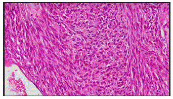
The MRI of the Lumbosacral spine showed a round iso-intense lesion in the left psoas muscle which was brilliantly hyper intense after contrast uptake. We operated the patient under general anesthesia in rt. lateral position. We approach the tumor through left retroperitoneal plane and found a round shaped, capsulated tumor along the distribution of ilio-hypogastric nerve which was excised. The excised tumor was firm, yellowish, capsulated and well circumscribed. It was attached with a nerve which had come out through the L2 intervertebral foramen. The specimen was sent for histopathological study which revealed a benign schwannoma. (Figure 1-3).
Figure 1: T1 contrast MRI Coronal and axial section at the level of L4/L$ showing the tumor in the left psoas muscle.
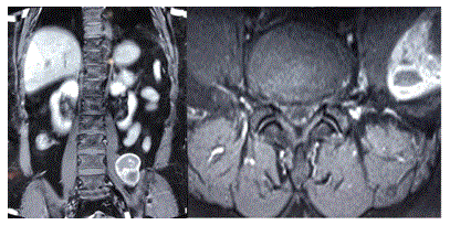
Figure 2: intraoperative view of the tumor.
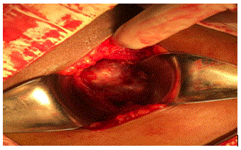
Figure 3: The whole tumor after resection.
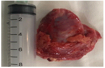
Figure 4: Histopathological photomicrodraph (H&E stain).
Discussion
Retroperitoneal schwannomas can be excised by retroperitoneal approach. Though it is also removed by use of laparoscopic approach [8]. It is very convenient but there may be difficulties if the tumor was attached to the major vessels8. Preoperative establishment of diagnosis is difficult in case of retroperitoneal schwannomas, however close relationship of retroperitoneal tumors with adjacent neural structures in imaging studies should raise a suspicion. Complete surgical resection is the treatment of choice. Histology and Immunohistochemistry confirms the diagnosis2. In our patient, during surgery the tumor had come from the left iliohypogastric nerve and the histopathological examination had confirmed it as schwannoma.
The retroperitoneum is flexible and nonrestrictive; therefore, a large, deeply situated tumor is usually present before patients have any symptoms These masses tend to displace retroperitoneal structures such as the kidney and ureter but generally do not invade them [9]. In one series the size varied from 8.8 to 20 cm in the greatest diameter9. Our tumor was also about six cm in largest diameter. Retroperitoneal schwannoma can present with varied symptoms. They mimic as a psoas abscess [10], ureteric obstruction [9], vague abdominal pain [11] or neurological problems as in our patient. Almost all schwannomas show intense immuno histochemical staining for S-100 protein, confirming the neuroectodermal origin of the tumor cells [4].
Furthermore, because even in benign schwannomas a local recurrence rate of 10-20% has been described, probably due to incomplete surgical excision, a more conservative approach such as enucleation is not sufficient [4]. Therefore, complete excision is the surgery of choice. In our patient we had excised the tumor along with part of the healthy nerves on either side. The retroperitoneal approach affords direct access to lumbar paraspinal lesions. There are two other possible approaches. One is a trans peritoneal approach via the midline, and the other a posterolateral approach through the Sacro spinalis muscle. The former carries the risk of damaging the inferior vena cava and ureter. The latter has the disadvantage of severe postoperative muscle pain which may persist long after surgery [12]. Therefore we had chosen the retroperitoneal approach. The approach was straight forward and the healing was satisfactory.
Conclusion
Psoas schwannoma is a rare tumor. It may present with different presentations. Abdominal ultra-sonogram can determine its presence but it can be confirmed by abdominal MRI. Retroperitoneal approach and total excision can be carried out with no or minimum morbidity.
References
- Fu C-Y, Lin C-H, Yu T-C, Lu T-C, Hsieh C-B, et al. (2008) Solitary benign schwannoma of the psoas muscle: A case report. Visceral Medicine 24: 249-251.
- Theodosopoulos T, Stafyla VK, Tsiantoula P, Yiallourou A, Marinis A, et al. (2008) Special problems encountering surgical management of large retroperitoneal schwannomas. World Journal of Surgical Oncology 6: 107.
- Claes H, Oyen R, Stessens R, Vereecken R (1987) Solitary benign schwannoma in the psoas muscle. The journal of Urology 137: 753-756.
- Tortorelli AP, Rosa F, Papa V, Rotondi F, Sanchez AM, et al. (2007) Retroperitoneal schwannomas: diagnostic and therapeutic implications. Tumori Journal 93:312-315.
- Kuyumcuoglu U, Germiyanoglu C (1990) Solitary benign schwannoma in the psoas muscle. International urology and nephrology 22:107-111.
- Loke T, Yuen N, Lo K, Lo J, Chan J, et al. (1998) Retroperitoneal ancient schwannoma: Review of clinico-radiological features. Australasian Radiology 42: 136-138.
- D'Silva KJ, Dwivedi AJ, Barnwell JM. (2003) Case report: Schwannoma of the psoas major muscle presenting with abdominal and back pain. Digestive diseases and sciences 48: 1619.
- Seo IY, Boldbaatr Y, Choi KH (2011) Laparoscopic Resection of Ancient Schwannoma Embedded in the Psoas Muscle. Surgical Laparoscopy Endoscopy & Percutaneous Techniques 21:e336-e338.
- Daneshmand S, Youssefzadeh D, Chamie K, Boswell W, Wu N, et al. (2003) Benign retroperitoneal schwannoma: a case series and review of the literature. Urology 62: 993-997.
- Liu Y-W, Chiu H-H, Huang C-C, Tu C-A (2007) Retroperitoneal schwannoma mimicking a psoas abscess. Clinical Gastroenterology and Hepatology 5: A32.
- Makhoul E, Kamel R, Hanna N (2017) Schwannoma of the psoas: An unusual cause of abdominal pain. Arab journal of gastroenterology 18: 44-46.
- Hida K, Iwasaki Y, Abe H, Itamoto K, Kaneda K, et al. (1993) Schwannoma in the psoas muscle removed by the retroperitoneal approach. British Journal of Neurosurgery 7: 213-215.