New Developments in Neuroscience. A Review
Aage R. Møller1*
1 The University of Texas at Dallas School of Behavioral and Brain Sciences, USA.
*Corresponding Author: Aage R. Møller, The University of Texas at Dallas School of Behavioral and Brain Sciences, USA, TEL: 972-883-4306 ; FAX: 972-883-4306;E-mail:amoller@utdallas.edu
Citation: Aage R. Møller (2019) New Developments in Neuroscience. A Review. Archives Neurol Neurosurgery 3:113.
Copyright: : © 2019 Aage R. Møller. This is an open-access article distributed under the terms of the Creative Commons Attribution License, which permits unrestricted use, distribution, and reproduction in any medium, provided the original author and source are credited
Received date: January 18, 2019; Accepted date: January 28, 2019; Published date: February 01, 2019.
Abstract
Many more people are now engaged in neuroscience research than just a few years ago, and the availability of new technologies has now made it possible to do studies that were not possible a few years ago. Recent studies have brought a new understanding of the normal functions of the nervous system it has made it possible to study diseases nervous system in new ways.
New techniques have made it possible to study morphology and function of the nervous system in healthy humans and individuals with diseases. The finding that the brain is a distributed system with extensive and dynamic functional connections (connectivity) between different systems has brought new understanding of the role of functional connections and neuroplasticity in creating symptoms and signs of diseases has been explored.
Earlier it was believed that age-related changes in the brain included loss of nerve cells. Recent studies have shown no significant change in the number of nerve cells, but there is a loss of anatomical connections and both increases and decreases in the strength of connections. The progression of the age-related changes has great individual variations but in general follow behavioral decreases in visual retention abilities as indicated by the results of the Benton test.
Recent studies have found that activation of maladaptive neuroplasticity plays the primary role in initiation and continuation of diseases such as chronic neuropathic pain, spasticity, severe tinnitus and it is an important cofactor in many other neurological disorders. That activation of neuroplasticity may control the immune system was an important finding that has had practical clinical importance. Increased understanding of the role of stress in the risk of many kinds of diseases together with maladaptive neuroplasticity likewise plays a central role in creating and in the maintenance of these diseases has opened new possibilities for treatment. These results have suggested new ways to reduce the risk of acquiring many diseases beyond neurological diseases including infectious diseases and cancer.
The finding that activity in the afferent part of the vagus nerve can reach many parts of the brain through the nucleus of the solitary tract has opened possibilities of affecting many functions through vagus nerve stimulation and possible treatments of neurological diseases.
This review will focus on systems neuroscience and describe and discuss some of the advances in the understanding the biology of the central nervous system and the clinical implications of that knowledge.
Introduction
The brain is a giant information processor that can extract useful features in many forms of sensory signals. The brain is also a controller of complex motor systems and the site of human creativity and consciousness. It has enormous memory capacities, estimated to be in the size of 2.5 petabytes (2,500 terabytes) (Reber, 2010). The human brain has approximately 100 billion (1011) nerve cells; weight 1.3-1.4 kg, and in early pregnancy, the rate of cell growth is approximately250,000 cells/minute. At rest, the brain uses about 20% of the body's energy.
The human brain is the most complex structure known to man. The complexity of the brain can compete with that of the universe. Neuroscience, the study of the brain and other parts of the nervous system, developed first slowly and then at a rapid pace. Similar to research in many other areas, people's curiosity and ingenuity drive research in neuroscience.
Models of the neurobiology of the brain have traditionally been viewed as having two main parts, one regarding how the brain is organized, how different systems work and interact. This is known as systems neuroscience. The other branch of neuroscience is the study of biochemistry, now often referred to as molecular biology. That branch of neuroscience is concerned with the structure and the working of the elements of the central nervous system, the nerve cells and other cells that are important for the function of the central nervous system.
There has recently been tremendous progress in understanding the structure and function of both of these goals of a study of the central nervous system. First, neuroscience studies have aimed to describe structure (anatomy) and later to understand functions. Recently the third goal of neuroscience research has emerged namely the use of basic scientific knowledge and understanding to design methods for treatment of diseases and for reducing the risk of acquiring neurological diseases. Several different parts of the brain do the same task.
Early most studies of the nervous system were done in animals, but new technology and techniques have gradually opened possibilities to do physiological studies in humans. Studies in patients undergoing neurosurgical operations started with Wilder Penfield (1891-1976), has made it possible to study both the normal and the diseased nervous system.
The hypothesis that the different parts of the brain do not operate independently, and many parts of the brain participate in many functions has been supported by experimental studies in humans. Tasks, even simple ones, engage many parts of the brain simultaneously.
The general objective of neuroscience research has gradually changed. Earlier much importance was ascribed to the function of single nerve cells while no evidence shows that it is the concerted actions of populations of nerve cells that determine which features of the neural code in elements of the of neural networks in the brain. Earlier much importance was ascribed to the discharge rate, but the recent interpretation of the results of animal experiments have shown little importance of the impulse frequency of the neural activity in brain networks. Instead, there is now a general agreement among neuroscientists that the coherence of neural activity is the important features of the activity that controls awareness and the discrimination between different cognitive variables.
Earlier only the anatomical aspects of the connections could be studied, but now functional aspects can be studied. Such studies of functional connections in the CNS have developed into a subspecialty “connectivity” of neuroscience. Studies have shown that many anatomical connections usually are not functional. Functional connections exist between most regions of the brain, and the connections are often reciprocal, creating loops where information can circulate building the basis for iterative interpretation. The strength of the connections varies widely through the activation of neuroplasticity. Neuroplasticity thus makes many functional brain connections dynamic. Extensive anatomical connections exist between most regions of the brain, and the connections are often reciprocal, creating loops where information may circulate. The different parts of the brain interact through reciprocal (two-way) connections between different parts. Many parts of the brain are now regarded to be involved in everyday tasks, and some parts of the brain can do more than one task. Brain systems may be similar to the hubs of the airlines in the USA. The functional connections in the brain are dynamic. Not all connections are functional because of ineffective synapses.
Changes in connectivity have been related to symptoms and signs of disease such as chronic neuropathic pain and severe tinnitus and including age-related symptoms and signs. There are many similarities between for example chronic neuropathic pain, severe tinnitus and age-related symptoms and signs [1].
The results of studies of the functional organization of neural network in the brain have brought new understanding of many functions and studies of deviations from the regular organization in diseases has brought new understanding of the causes of the symptoms and signs diseases. There is a consensus that network dysfunctions are related to many disorders of memory including age-related diseases such as dementia [2].
History of the development of modern neuroscience
Our understanding of how the brain is organized has changed over time; First slowly, then at a rapid pace. Galen (Aelius Galenus or Claudius Galenus, AD 129 – 199 or 217) dominated medical education from about year 280 until the year 1543 (about 1250 years). His writings and teaching were based on dissection of pigs and monkeys. During that time, it was not permitted to dissect humans. Galen also presented proof that the brain controls the motion apparatus. Over a twenty-five-year period Leonardo da Vinci (1452-1519) dissected about thirty human corpses but in the year 1515 the Pope, Leo X, orders him to stop. He contributed a series of anatomical drawings based on autopsies of humans. The accuracy of his observation was better than anything previously attempted. Cartesius (Descartes) (1596-1650) a mathematician and a physiologist and much more contributed to mathematics and gave the name to the common (Cartesian) coordinate system. His dualistic principle of sensorimotor processes consisting of a system that is predictable (mechanistic) and one that is not (the soul) was the basis for discussions by many scientists for many years to come, and it is still valid. He used the withdrawal reflex as an example of functions that could be replicated by mechanical devices, but he assumed the soul was complicated and located in the pineal gland (he chose the pineal gland because it is a single structure).
The birth of experimental neuroscience
It was not until experimental neuroscience was born that important questions could be answered in neuroscience. That more or less started with the work of Francis Bacon (1561-1626) who is credited by having introduced experiments in studies of biology. He advocated a critical view on old science, was arrogant and convicted of several crimes including bribery. Francis Bacon had many important positions in the parliament and the government but is mostly remembered for his philosophical writings. Many years later Charles Scott Sherrington (1857-1952) did experimental work that meant a major increase in knowledge about the biology of the brain. He is regarded as the father of modern neurophysiology for his work on communication between neurons through synapses. Specifically, he studied spinal reflexes and pointed to the importance of the interaction between different parts of the brain.
There were many other important discoveries from late 1700 to the present time. Francois Magendie (1783-1855), discovered that sensory information enters in dorsal spinal roots while motor information exists in ventral roots, Ivan Sechenov (1829-1905), Russian physiologist whom 1863 discovers inhibitory synapses. The Hungarian anatomist J. Szentágothai (1912-1994), described the structure of the synapse using the elctronmicroscope that was just invented. John Eccles, (1903-1997), an Australian neurophysiologist who studied synapses and the somatosensory system. He became generally known for his philosophical views on the "brain-mind problem." Eric Kandel (1929-), Austrian born American medical doctor who specialized in Psychiatry and became a prominent neuroscientist won the Nobel Prize for studies of the basic function of synapses in memory storage.
Some of the founders of modern neuroscience have asked some critical questions regarding the possibility to emulate the functions of biological systems, and this is still an important part of the human activity to find ways to replicate normal biological functions. One of the first attempts to imitate functions of animals by mechanical devices was the Vaucanson's duck, displayed in the gardens of the Tuileries, France, 1738 and designed by a 29-year-old watchmaker, Jacques de Vaucanson. Vaucanson's duck may be regarded as the first attempt to make a robot. More recently many computer models have been proposed for emulating specific functions of the nervous system.
Brain structures that have gained recent attention
The insular lobe and the vagus nerve are two structures that have received much attention recently.
The insular lobe
The insular lobe is the fifth lobe of the brain. It is located deep under the temporal love, and it is not visible from the surface of the brain. The function of the insular lobe has been little known until recently. The insular cortex is often the site of epileptic foci, and a new diagnostic method for localizing foci of epileptic seizures have provided some valuable information about the various functions related to the insular lobe. The method using recordings from many sites in the insular lobe for determining the anatomical location of foci of epileptic seizures [3] has yielded much new functional information about the function of the insula. The results of studies of 5 patients who reported viscerosensitivity in one part of the insula and somatosensitive reactions upon stimulation at an anatomically different part of the insula with stimulations of one at a time of the 113 electrodes that were inserted in the insula [4]. The electrodes were implanted during anesthesia, and the patient is kept in the hospital for several days while the different anatomical locations are tested with the patient awake and cooperating.
The fact that patients undergoing such diagnostic studies are conscious and able to communicate what they experience when specific anatomical locations are stimulated yielded valuable results regarding the function of the structures of the insular lobe. The results of these studies have shown that the insula may be the site of many common functions and some unexplained symptoms of neurological diseases may originate in the insula. These studies of the functional properties of the insular lobe are just one example of how valuable modern diagnostic methods can be beneficial outside their original field of use.
There is also recent evidence that the insular lobe, is involved in several neuropsychiatric diseases [5] and goal-directed cognition, conscious awareness, autonomic regulation, interoception, and somatosensation and migraine [6]. The insula has extensive connections with many structures in the brain. Activation of these structures is at least partly responsible for the distress and mood symptoms such as depression that often accompany disorders of phantom sensations.
The insula and the nuclei of the amygdala and the anterior cingulate are now known to be extensively involved in diseases, such as chronic neuropathic pain and some forms of tinnitus, and disorders that involve parts of the old brain such as the nuclei of the limbic system, also known as the emotional brain [] (LeDoux 1996), in addition to sensory cortices.
The vagus nerve
The vagus has been known for supplying parasympathetic innervation to the heart, the vocal cords and organs in the lower abdomen including the reproductive organs. These efferent fibers occupy only 20% of the nerve fibers of the vagus nerve and the remaining 80% are afferent fibers that innervate sensory organs in the same organs that the efferent fibers innervate, but the function of afferent fibers have only recently gained attention from neuroscientists. The cells in the nucleus of the solitary tract (NST) which are the target of the afferent fibers of the vagus nerve sends axons to many parts of the brain (Figure 1), which means that the vagus nerve can influence many organs in the abdomen can influence many brain functions.
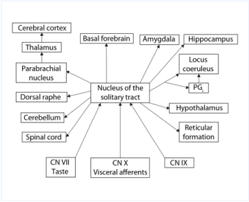
The vagus nerve through the cells in the NST can influence the cholinergic system of the forebrain (the nucleus of Meynert) [7,8]. The stimulation of the vagus nerve can, therefore, promote activation of neuroplasticity and that may be used to reverse the adverse effects of maladaptive plasticity thus alleviating the symptoms and sign of some common diseases [9,10].
Recent studies have shown that the vagus nerve plays a vital role in memory and neuromodulation by vagus nerve stimulation may, therefore, be used to treat disorders of memory through connections from NTS through the locus coeruleus to the hippocampus (McIntyre, 2012).
Recent progress
Recent progress in studies of the anatomical organization of the central nervous system
New techniques make it possible to study the architecture of both white matter (nerve fiber tracts), and gray matter (clusters of nerve cells) [11]. Earlier techniques for morphological studies of structures of the central nervous system staining and light microscopy produced just a two-dimensional view. New techniques, known as diffusion tensor imaging (DTI) has become one of the most popular MRI techniques in brain research, as well as in clinical practice [12]. It is used to study white matter architecture in many different diseases. Tractography is a 3D modeling technique using data collected by diffusion MRI to represent nerve tracts visually. It uses special techniques of magnetic resonance imaging (MRI) and computer-based diffusion tensor imaging.
The diffusion tensor imaging (DTI) methodology that was introduced in the year 1994 has been used to study the white matter architecture and integrity of the brain of people with diseases such as in multiple sclerosis, stroke, aging, dementia, schizophrenia, and more [13].
Recently it was shown that the use of chemical treatment with a kind of detergent can remove surrounding material make a specimen of the brain transparent by removing surrounding material. This technique known as CLARITY together with fluorescent labeling (by Karl Deisseroth) can make nerve cells in an intact mouse hippocampus visible [14,15]. These new techniques produce beautiful pictures that may provide information not obtainable with other techniques.
Biochemistry of the brain
Neural receptors and transmitters have been studied extensively, and the Nobel prize has rewarded many of the major discoveries in neuroscience. Julius Axelrod, Bernard Katz, and Ulf von Euler are known for fundamental work on work on neurotransmitters and Alfred G. Gilman, and Martin Rodbell discovered G-protein-coupled receptors and their role in signal transduction. Arvid Carlsson, Paul Greengard, and Eric Kandel made fundamental discoveries concerning chemical signal transduction in the nervous system.
Functional organization of the brain
Functional connections or functional networks of the brain have been studied extensively recently. The techniques for doing that are derived from statistical descriptions of time series data, such as in resting-state functional MRI (fMRI) studies or from time series analysis of the different oscillations in the EEG. These methods make it possible to determine the strength of the different connections. Other technologies such as the magnetoencephalography (MEG), processing of EEG using time series analysis have and analysis of functional MRI have made it possible to identify which anatomical connections are functional, and the strength of the connections and its changes can now be studied using techniques described by Schlee and his co-workers [16,17]. MEG is one of the most accurate methods to determine functional connectivity, and it makes use of measurements of the extremely small magnetic fields generated by the electrical activity in the brain.
Computational processing of EEG recordings makes it possible to determine the strength of the individual connections between different parts of the brain. Such studies performed in animals and humans have yielded a welch of information about the normal function of many brain structures as well as how functions are altered in many neurological diseases.
Recording of the Gamma component of the EEG (40-100Hz), is used as a measure of functional cortical activation. Gamma activity is assumed to be implicated in creating the conscious perception of sensory signals in the cerebral cortex although there is still some uncertainty regarding the actual meaning of these particular brain rhythms [18]. Some of the early attempts to study the functional connectivity in the brain used what was known as resting-state fMRI using the blood-oxygen-level-dependent (BOLD) signal. That method is still in use [19].
The recent evaluation of the functional organization of the brain has revealed that large-scale brain networks consist of multiple segregated subnetworks of interacting brain areas [20]. Descriptions of resting- state network architecture have provided clues for understanding the functional significance of the different parts of these segregated subnetworks.
The functional organization of the brain cannot be inferred from the results of morphological studies because the functional connections depend on the efficacy of the synapses that connect the axons to their target cells. Recently a wealth of observations that have been made of human brain networks defined by resting-state functional correlations (RSFCs) of BOLD time-series have been published. Analysis of these observations has brought new understanding of neural networks in the brain and their wide interactions [20,21]. In these studies, it has been regarded that the brain consists of a series of complex neural networks that are interconnected through many paths.
Combining data from numerous empirical and computational studies, network approaches strongly suggest that brain hubs play important roles in information integration underpinning numerous aspects of complex cognitive function [22,23]. Network theory identifies several highly connected and highly central hub regions and predicts that these network hubs and their connections play key roles in the integration of information and in achieving efficient neuronal signaling and communication in the brain.
Network analysis tools applied to structural and functional human connectome data provide a data-driven computational framework for detecting brain network hubs and for examining their variation across individuals, their development across time, and their roles in brain disorders [22].
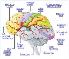
The brain has earlier been regarded to be a compartmentalized system of a series of structures with specific functions as illustrated in Figure 2, but current concepts regard the brain to be more like a distributed system.
It is now generally accepted that the brain is a distributed system where many functions involve many different parts of the brain.
It has been known for a long time that the brain is plastic (malleable) for the most part, but a new understanding of many details has emerged recently [10]. For example, it is well-known that activation of neuroplasticity can change many functions whereas other functions such as long-term memory, sexual preference, personality, and handedness seem to be "hard-wired." Many studies have confirmed that maladaptive plasticity plays a vital role in common diseases such as chronic neuropathic pain, spasticity, and probably also disorders such as fibromyalgia.
There is evidence that the normal connections between different parts of the nervous system changes constantly and that altered connections are important features of the symptoms and signs of many neurological diseases. Earlier the symptoms and signs of neurological disorders were assumed to be related to malfunctions of neural circuits while there are now indications that altered connections may play a more important role in abnormal neural functioning including that of aging [2,16,24].
Quantitative studies of connectivity in persons who do not have symptoms of a disease have provided new understanding of the functional organization of the brain and quantitative studies of the functional connections in the CNS is becoming a new area in neuroscience known as "studies of connectivity." Results of such studies have confirmed that the brain is a distributed system where there are functional connections between many parts of the brain and the spinal cord. Most of the connections are reciprocal (two-way) creating loops where information can circulate providing possibilities for iterative analysis of information.
Functional connections in the brain may have a similar structure to modern communication systems such as the route map of a major airline with a hub and spoke structure [25]. Like an airline's service to the different cities can be measured as the number per day of flight from a hub, the strength of neural connections from one structure of the brain to other structures have different strengths as measured by MEG or EEG recording. In the brain, the strength of connections is a function of the number of axons and the efficacy of the synapses that connect the axons to their target cells. The number of axons is fixed, and the efficacy of the synapses that connect the axons to their target cells may change through activation of neuroplasticity.
The resting state connectivity is related to cognitive changes [22] and there is evidence that virtually all domains of cognitive function require the integration of distributed neural activity. These investigators found that network analysis of human brain connectivity has consistently identified sets of regions that are critically important for enabling efficient neuronal signaling and communication.
Recent studies of the functional connections in the brain together with the use of modern neuroanatomical studies have the organization of the brain can change during life and what the results of age-related changes may affect a person [20,21,26].
Studies of connections in the brain have helped to explain many features of diseases. For example, it has recently been shown that the parahippocampus is involved in multiple networks that are active in different forms of expression of diseases such as neuropathic pain and tinnitus [27,28]. Thus, a single hub can be involved in multiple overlapping networks (Figure 3).
Mapping of the cerebral cortex
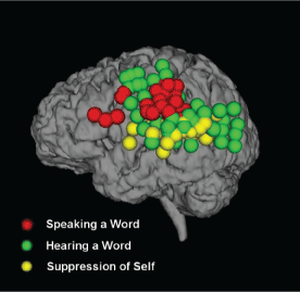
A certain task activates larger parts of the brain that was earlier believed. Production of speech and interpretation of spoken words were earlier believed to involve only Wernicke’s and Broca’s areas of the brain (Figure 4). Recent studies have shown that large parts of the brain are involved in the interpretation of spoken words and that has practical importance for neurosurgical procedures where parts of the cerebral cortex are resected. Mapping the cerebral cortex (electrocortigography, ECoG) for gamma activity (EEG activity in the frequency range of 40-80 Hz) has shown indications that the perception of a spoken word involves larger areas of the cerebral cortex than what was earlier believed [29] (Figure 4).
These studies made use of recordings of the gamma component of EEG recordings, obtained from the exposed dura mater in a patient undergoing various kinds of neurosurgical procedures. The gamma waves include a spectral component in the frequency range of 40-100 Hz. These components are regarded to be an indicator of functional activity in the cerebral cortex. These high-frequency components are attenuated by the scalp and skull bone and can therefore not be studied using traditional EEG recordings from electrodes placed on the scalp.
There are many other examples of neural systems that show that different tasks can be performed in the same part of the brain and different kinds of processing can be performed in the same structure. For example, pain and addiction share the same brain regions [30].
Widespread gamma activity was observed during the presentation of words (green). When the patients repeated the words, activation was primarily observed over frontal and parietal cortex (red). The location of the electrodes that were used to record the gamma activity are indicated by yellow that responded to hearing the words when presented by the computer, but not when the patient repeated the word (yellow). The results shown in Figure 4-6 were based on composite data from 12 participants.
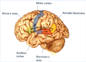
Figure 4: Classical description of the brain regions that are primary to hearing and generation of speech sounds.
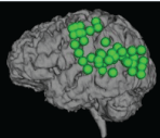
Figure 5: Gamma activity in awake persons who were asked for the meaning of spoken words [29]. Add to the figure legend: Reproduced from: VI Towle, et al. (2008) ECoG Gamma Activity: Differentiating expressive and receptive speech areas. Brain, 131: 2013-2027, with the permission of the Publisher.
The control of body functions by the mind (“thinking”)
It is not only sensory signals that can affect body functions but also the "mind" can control many functions, such as how muscles contract. Recent studies have revealed that the “mind” can influence muscle control. Figure 7 shows an example of how just thinking can alter the strength of contraction of muscles of the hand elicited by stimulation of the motor cortex. Figure 7 shows an example of how just thinking can alter the strength of contraction of muscles on the hand elicited by the same electrical stimulation of the motor cortex.
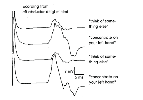
Many other functions can be voluntarily manipulated by the mind such as the effects of stress on body function can be affected by the mind. This means that it is not only external (sensory) signals and internal signals from body organs in the abdomen, but also voluntary and involuntary signals from the brain can affect basic body functions. The EMG activity from muscles of the hand shown in Figure 7 was elicited by electrical current in the motor cortex induced by magnetic impulses applied through the skull to the brain. The method of transcranial magnetic stimulation (TMS) is a practical way to activate brain structures by inducingan electricall current in the brain. It is painless and does not cause any other noticeable inconvenience.
Development of the brain
It has been confirmed that that the development of the brain is extensive during the first two-three years of life and it continues at a slower speed until the age of twent and before birth the rate of cell growth in early pregnancy is 250,000 cells/minute [32].
Postnatal development
It has been shown recently that the brain undergoes major changes during the first two years of life with substantial pruning of connections and programmed cells death is extensive during that period. The changes in the brain continues to the age of 20. Before birth (20wk gestation) there are more than 1,000,000 cells per mm3 in the visual cortex. At birth that has been reduced to approximately 90,000 cells per mm3, at the age of month it is further reduced to 40,000. This number stays unchanged tothe agee of 80 years. The average number of synaptic contacts on a cell in the neocortex for a newborn was found to be approximately 1,500, in early childhood, approximately 15,000, and in adulthood the average number of synapses per cell has increased to an average of 7,500 [32].
Age-related changes
It was earlier believed that loss of nerve cells in the brain was the cause of the memory loss that is common at old age but it has recently been shown that there is little to no loss of nerve cells in the brain of an older individual [32]. While it has been confirmed that the brain shrinks with age, it is not the nerve cells that are lost as was earlier believed but it was a loss or a shrinkage of other kinds of cells that makes the brain shrink. The age-related shrinking of the brain must, therefore, have other causes than nerve cell loss. Loss or shrinking of glial cells may be one of the causes of the shrinking of the brain that usually occurs with aging.
| Nerve cells | Glial cells | |
|---|---|---|
| 65-75 years old | 17.9x10⁹ | 41x109 |
| 76-85 years old | 18.1x10⁹ | |
| 94-105 years old | 16.32x10⁹ | 29x109 |
Data from [33].
While the average number of nerve cells remains rather stable during adult life, at least until 80 years of age, there exists a considerable loss of nerve fibers in the brain late in life [34]. The estimates of the total length of axons, in the brain in kilometers for males at the age of 20 years was found to be approximately 176,000 km, and it has decreased to 97,000 km at 80 years of age. The total length of the fibers in the brain of females at the age of 20 years was found to be 149,000 km, and at the age of 80 years, it was 82.000 km [34].
Neuroplasticity
Recent studies have brought much interest in the role of activation of neuroplasticity in normal neural functions and in the role of neuroplasticity in diseases [10]. The concept of neuroplasticity has changed; earlier it was believed that the brain and the spinal cord were finished developing at birth. Then came a time when it was believed that all neural functions could be changed by activating neuroplasticity. More recently it was confirmed in many studies that the brain is plastic for the most part and that some functions are harder to change than others and that there are functions that are very hard to change [10]. Long-term memory depends on neuroplasticity but cannot be changed. Some functions are stable from birth and cannot be changed. Sexual preference, handedness, personality are functions that have been subjected to many attempts of changing but with little or no success.
Activation of neuroplasticity makes it possible to adapt to changing demands and neuroplasticity is necessary for normal childhood development. Both beneficial and harmful plasticity play important roles in the recovery after stroke and traumatic brain injuries. Activation of neuroplasticity is the basis for learning new skills and memory functions, but recent studies have revealed that activation of neural plasticity also plays roles in initiating symptoms and signs of some neurological diseases. Activation of neuroplasticity alters neural function mainly through changes in synaptic efficacy that can alter the strength of connections in the CNS. While many forms of activation of neuroplasticity are beneficial neuroplasticity may also be associated with the non-beneficial effect such as creating symptoms and signs of disease. This kind of neuroplastic changes is known as maladaptive neuroplasticity.
Maladaptive neuroplasticity is the primary factor in the creation of specific disorders, such as chronic neuropathic pain [10], spasticity, severe tinnitus; moreover, activation of neuroplasticity plays crucial roles in many other neurological diseases such as the symptoms and signs of spinal cord injuries (SCI) [35] and ischemic strokes. This is one of the dark sides of neuroplasticity [10]. Activation of other forms of neuroplasticity is driving the recovery of stroke, and other forms of traumatic brain injuries (TBI) and spinal cord injuries (SCI).
The role of neuroplasticity in the development of the brain
It has been known for many years from studies in animals such as the classical study of vision by Wiesel and Hubel [36] that activation is essential for the normal development of the nervous system, anatomical as well as functional [36]. Subsequent studies have confirmed that the absence of activation of neuroplasticity in the brain of young children may have serious consequences.
Recent animal studies indicate that unused regions of the brain can be taken over by other systems. It was demonstrated in animal experiments that the input from the visual system could take over unused areas of the hearing cortex [37,38]. This miswiring of the brain caused cells in their auditory cortex to respond to light stimulation because of the misdirected visual information. These replacements may be permanent. This is one reason why it is essential that babies who are born with sensory deficits such as hearing loss get some input to their auditory nervous system early in life.
Activation of harmful neuroplasticity
Activation of harmful plasticity (maladaptive plasticity) can cause or promote diseases such as chronic neuropathic pain, severe tinnitus, spasticity and some forms of muscle spasm ("Plasticity diseases.") Other factors such as stress amplify the risk of getting one of these diseases. The phantom limb syndrome is the creation of phantom sensations (pain and tingling) after amputation of a body part (a limb). Activation of neuroplasticity is involved in some age-related changes.
The value
The value of preventive means in medicine has been re-evaluated
Earlier the focus in medicines has been on finding diseases early and providing diagnosis and treatment of the diseases. Recent development in clinical medicinehase provided a recognition of prevention (reducing the likelihood of getting a specific disease) and a more realistic view on the value of routine check-ups in people who have no symptoms of a disease or no hereditary indications of specific diseases. The value of some routine check-ups has been disputedfromf recent studies [39]. Many people will worry for the next check-up because check-ups only concern the history of a person’s medical condition. It has been shown that many commonly used routine check-ups have false positive tests that may cause treatments of diseases that person does not have, and that tests can have risks such as from ionizing radiation, which increases the risk of cancer. The ionizing radiation used in theconventionaln mammography has been estimated to induce cancer in one of 1,000 women who have mammograms over a lifetime [40].
The increase in knowledge about how to reduce the risk of many severe diseases and the increased availabilities of preventive means have made it possible for many people to live a better life by reducing the risk of diseases. Vaccinations are now available for many diseases, and this can save a person’s lif, and it is an effective way of reducing the risk of many severe infectious diseases. However, many people ignore to use these very effective and beneficial means, a problem that has attracted little research. That means that the real problem is to convince people of the importance of these simple precautions. Recent research in neuroscience has emphasized the importance of a person's lifestyle for longevity and quality of life. Physical exercise has many positive effects. It increases the production of BDNF and, more recently, it was found that physical exercise also facilitates the recycling of organelles in nerve cells, another example of how extensive and complex interactions between widely different body and brain systems are. Minor changes in lifestyle may be sufficient to delay the onset of dementia and Alzheimer's disease, and perhaps age-related hearing loss [41].
The value of vitamin D3 as a supplement has been emphasized by recent studies that have shown that daily intake of vitamin D3 (4,000IU daily) can improve health especially reducing the risk of severe diseases such as cancer and boosting the immune system and reducing the effect of oxidative stress has become evident recently [42]. Recent studies have estimated that the use of sunscreen that is effective in reducing the risk of skin cancer but deprive a person of the synthesis of Vitamin D3 that is effective in reducing the risk of cancers that have less effective treatment than skin cancers and poor prognosis. Most of a person's vitamin D3 comes from exposure to sunlight and, therefore, lack of exposure to the skin of sunlight. This can be caused by wearing clothing or when a person is indoor or using of sunscreen. Lack of vitamin D3 have an adverse health effect [43] which can be restored by taking a supplement of vitamin D3. It has been estimated that 45,000 people die every year in the USA from cancers that were caused by a lack of D3 vitamin [43].
Congenital disabilities are costly, and cause loss of quality of life. Recent studies have shown that the risk of many congenital disabilities can now be reduced. That means nearly 120,000 babies are affected by congenital disabilities each year which means that one in every thirty-three babies born in the United States each year has a congenital disability.
Spina bifida is one of the most common congenital disabilities, the occurrence of 1.4 per 1,000 pregnancies. It is a developmental disorder that causes severe disability because some of the vertebrae are not fully formed during before birth. After birth, these vertebrae are not correctly connected and open. If the opening is large, a portion of the spinal cord can protrude through the opening. Surgery can not sufficiently repair the defects, and people with spina bifida have severe signs such as leg weakness, hip dislocation, scoliosis, bladder and bowel problems, urinary tract infections, reduced kidney function).
Different studies have found substantial benefit from taking folic acid. If the mother takes a folic acid tablet before the start of pregnancy and every day the risk of giving birth to a baby with spina bifida is much reduced, some studies find a reduction of nearly 100%. (Centers for Disease Control and Prevention. Use of Folic Acid for Prevention of Spina Bifida and Other Neural Tube Defects 1983-1991). Spina bifida cases declined up to 70% in 2002. Taking folic acid before and during pregnancy can also reduce the risk of giving birth to children with cleft palate and anencephaly [44]. Other studies [45] found a 50% reduction in the incidence of anencephaly. More recently, it was shown that the risks of autism could be reduced by as much as 50% if the mother took folic acid before and during pregnancy [45,46].
However, only a few people take advantage of these possibilities of improving health and reducing the risk of acquiring severe diseases. It is surprising why this simple and inexpensive supplement that has no known side effects is not more widely used [47]. Now more than 35 years after the finding that taking folic acid could reduce the risks of giving birth to a child with spina bifida was published a study showed that only 35 % of mothers had taken folic acid before and during pregnancy [46].
It is not known precisely what leads to the development of autism and neural tube defects causing spina bifida, but it is assumed that it occurs very early in pregnancy, often before the woman knows she is pregnant. It is therefore essential to take actions that can reduce the risk of these congenital disabilities such as folic acid before being pregnant.
The cause of autism is unknown, but it has recently been shown that several factors are involved in the development of autism. There is evidence that air pollution, mainly by N2O, could increase the incidence of children born with autism. Exposure to agricultural pesticides during gestation is another factor that has been shown to promote the development of autism in the baby [41].
Intake of acetaminophen (paracetamol, commercial name Tylenol) is commonly recommended for young children for treatment of upper airway infections and other pain conditions, and for reducing fever; it is often added to other medications. Acetaminophen (brand name Tylenol) has been known for a long time to be hepatoxic in high dosages (4,000 mg per day), and it is the most common cause of liver failure.
Recently it was shown that it couldn’t have several other severe side effects if given to children younger than three 3 years of age [41,48,49]. These studies showed indications that acetaminophen can increase the risk of autism and possibly other diseases if given to young children. These findings were controversial but now based on several different several different studies. Connections in the brain are altered in individuals who have tinnitus. The auditory cortices are connected to other parts of the brain that generally are not involved in hearing.
Neural connections in diseases
It was earlier believed that symptoms of most neurological diseases were caused by pathology of specific parts of the nervous system and that symptoms are related to detectable morphological changes. It is now possible to explain some observations regarding neurological diseases using studies of connectivity and many recent studies have supported the concept that harmful neuroplasticity plays a more significant role than earlier assumed and there is evidence that the symptoms of many diseases are caused by changes in functional connections between parts of the central nervous system, many of which brought about by activation of maladaptive plasticity [50-52] Figure 8.
For example, recent studies have shown that the strength of the connections in the brain is different in people who have had tinnitus for a long time compared with that of people who have had tinnitus for only a short time [23,25].
Other studies have shown that the connections to the anterior cingulate and parahippocampus are stronger in persons who had a high degree of distress over a long time with tinnitus [51].
Brain networks
Brain networks involved in phantom perception
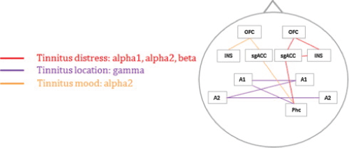
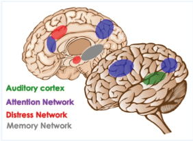
The symptoms of many neurological diseases are dominated by the perception of sensory signals that do not come from the outside, known as phantom sensations. The symptoms can be tinnitus, paresthesia, and different forms of neuropathic pain. Deafferentation playsessentialt roles in many diseases, especially chronic neuropathic pain, and some forms of severe tinnitus. Deafferentation caused by interruptions of peripheral nerves, or by fiber tracts in the brain. Maladaptive neuroplasticity activated by deafferentation is involved in creating the symptoms and signs of diseases such as chronic neuropathic pain, severe tinnitus, spasticit and the phantom limb syndrome. Many parts of the brain show functional changes in diseases where deafferentation is involved [50,52] Figure 9-11.
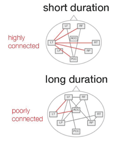
Figure 9: Functional abnormalities in the brain of persons with tinnitus are different for tinnitus of a short duration and tinnitus of a long duration. LF = Left Frontal, RF = Right Frontal, LT = Left Temporal, RT = Right Temporal, LP = Left Parietal, RP = Right Parietal, ACC = Anterior Cingulate Cortex, PCC = Posterior Cingulate Cortex. Reproduced by permission of the authors [51]. Reproduced from J Song et al. (2013) Distressed aging: the differences in brain activity between early- and late-onset tinnitus. Neurobiol Aging 34: 1853-1863 with permission from the authors.

Figure 10: Regions of the brain that are involved in diseases where deafferentation plays a primary role Add to the figure legend: Reproduced from B. Langguth et al. 2012 Neuroimaging and neuromodulation: complementary approaches for identifying the neuronal correlates of tinnitus. Front. Syst. Neurosci., after D DeRidder et al, (2011) Phantom percepts: tinnitus and pain as persisting aversive memory networks. Proc. Natl. Acad. Sci. U.S.A.108, 8075–8080, with the authors’ permission.
How do diseases such as chronic pain develop?
Many diseases have unknown causes, and it is not known how they start. There has recently been some progress in understanding how some neurological disorders start and it has been shown that stress is an essential factor in many neurological diseases. Stress affects both brain functions and body functions. It has developed for protection of the body and for adapting many brain and body functions to demands. Stress has manyadversee effects, and it causes many of its effects on body functions through the release of norepinephrine and corticosterone. Hans Selye coined the term "stress," and his students provided evidence that stress responses are related to the hypothalamic-pituitary-adrenocortical axis [53]. According to Selye’s “doctrine of non-specificity," stress is the nonspecific response of the body to any demand imposed upon it. Selye's "alarm reaction," the first stage of the General Adaptation Syndrome, would correspond to Cannon's "fight or flight" reaction.
Recent studies have confirmed that stress suppresses the immune system and thereby increase the likelihood of not only diseases such as virus infections but also cancer [41] and that stress also increases the risk of many kinds of cancer. Stress affects the cardiovascular system and promotes the development of hypertension, which is the primary risk factor for cardiovascular diseases and stroke. Recent studies have shown benefits of lowering the systolic blood pressure further than earlier recommendations (Group, 2015).
The increased release of epinephrine (adrenaline) and norepinephrine (noradrenaline) caused by stress promotes the conversion of acute pain into chronic neuropathic pain [54] (figure 12). These findings may lead to the development of methods for better pain control [30,55]. Figure 13.
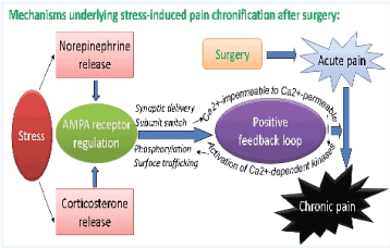
Figure 12: Mechanisms that are underlying a stress-induced process of transient pain progressing into persistent pain such as may occur after surgery. The model shows the role of stress on acute pain and in the development of chronic neuropathic pain. Courtesy Dr. Feng Tao, 2015 [54].
Stress also have positive effects, such as facilitating memory consolidation and retrieval [56].
Brain connectivity in ageing
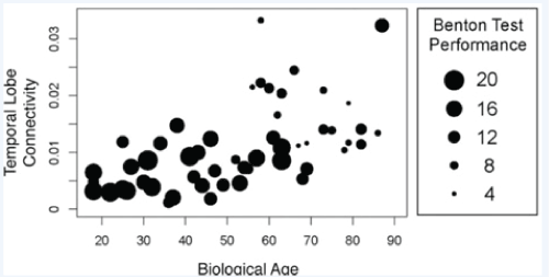
Recent studies have shown that the strength of some connections in the brain may increase, while the strength in other connections may decrease, as a person ages [26]. Age is associated with a reduced inflow to the medial temporal lobe system and a reduced input to the posterior cingulum/precuneous region of the brain. Age is also associated with stronger connectivity of temporal lobes, and that has been associated with decreased cognitive performance. There is a stronger inflow to the posterior region, and that is associated with better cognitive performance [16]. The changes in functional connections between specific parts of the brain are correlated with a decrease in the ability to retain visual information as shown from the results of the Benton test applied to people of different age.
Many forms of pathology in the body are followed by adaptive responses in the spinal cord and the brain. Damage to the facial nerve, for example, may be followed by functional changes in the facial moto nucleus causing such signs as synkinesis. Activation of neuroplasticity is a general pattern after injuries. These observations have a clinical implication because they make it possible to treat the synkinesis that of ten follows after the regeneration of peripheral nerves by appropriate physical therapy. Damage to sensory nerves can result in pain as a result of pathological alteration in pain circuits in the spinal cord, and the brain manifests as pain; damage to the cochlea can cause hyperactivity that is expressed in the form of tinnitus from changes in the function of CNS structures [57].
Effects
Effects on the nervous system from internal organs
Neurobiological role of the immune system
Interest in the immune system by neuroscientists had increased recently when it was found the nervous system controls many aspects of the immune system and that the immune system controls some aspects of the nervous system. The immune system is plastic (adaptive), similar to the nervous system. There are two kinds of immune systems: The innate immune system and the adaptive immune system. The innate immune system is programmed to respond to different kinds of intruders; it cannot "learn." The adaptive immune system is plastic and can "learn" (it responds stronger to repeating exposures). The brain and the spinal cord have their own immune system where microglia are first activated, then astrocytes.
New studies seem to indicate that the immune system is involved in creating diseases such as dementia and Alzheimer's disease. It has been shown that it is beneficial to reduce this reaction with medication such as minocycline [58-60]. There are also now indications that infections may play a role in degenerative diseases such as dementia. The vagus nerve plays a vital role with its release of acetylcholine; the cholinergic anti-inflammatory pathway is a neurophysiological route that regulates the immune system and the "vagal immune reflex." Inflammation can change the sensitivity of a peripheral nerve and its ganglion to mechanical stimulation. This is anessentialt factor in the pathology of main conditions such as low back pain [30].
Brain-Gut axis
The hypotheses regarding the relationship between the gut and the minddates back to the nineteenth century. The concepts of dyspepsia and neurasthenia gastric referred to the influence of the gut on human emotions and thoughts. The understanding of the importance of the brain-gut axis has recently been influenced by the attention that has been directed to the Helicobacter pyloris infection and its effect on diseases of the stomach and more recently its effect on many other systems [61].
It was earlier known that the brain may influence the functions of the gut through the vagus nerve, but it is only recently that it has been shown that the gut influences the function of the spinal cord and brain. This means that the brain-gut axis is a two-way control system. Information from the intestines exerts some control over the immune system, and the immune system has some control over body functions such as pain through the brain-gut axis. Bacteria in the gut produces a broad range of molecules that are neuroactive such as acetylcholine, catecholamines, gamma-aminobutyric acid, histamines, melatonin, and serotonin. These substances have many effects on the central nervous system. This flora consisting of probiotic, and pathogenic bacteria is established within the first few days of life, and it is maintained and modified during life. Recent studies show that bacteria, including commensal, probiotic, and pathogenic bacteria, in the gastrointestinal tract can activate the function of neural networks in the brain and the spinal cord. The results of such studies provide novel approaches aimed at affecting the function of the gut for the prevention and treatment of mental illness, including anxiety and depression [62]. The role of the gut flora on anxiety and mood disorders is now the subject of many animal and human studies [62,63].
The interest in the gut flora of bacteria was sparked by the study that showed that germ-free mice showed exaggerated HPA activity response to stress [64]; the fact that 70% of people with autism have gastrointestinal problems have been interpreted as a sign of involvement of the gut in the function of the brain [65]. A recent review of 25 animal and 15 human studies of the influence from the gut on central nervous system functions showed that probiotics could improve psychiatric disorder-related behaviors including anxiety, depression, autism spectrum disorder (ASD), obsessive-compulsive disorder, and memory abilities, including spatial and non-spatial memory [66]. Some of these substances can directly activate the vagus nerve [67-69].
Of pathological bacteria in the gut, Helicobacter pyloris stands out because of its high frequency and the cause of significant stomach pathologies ulcer, and stomach cancer. It was earlier believed that most of the common symptoms from the stomach such as gastric ulcer had anxiety and other mental reasons.
References
- Møller AR (2010) Similarities between tinnitus and pain In: Møller AR, Langguth B, DE Ridder D, Kleinjung T (eds.) Textbook of Tinnitus. New York: Springer.
- Dennis E, Thompson P (2014) Functional Brain Connectivity using fMRI in Aging and Alzheimer’s Disease. Neuropsychol Rev 24: 49-62.
- Stephani C, Fernandez Baca-Vaca G, Koubeissi M, Maciunas R, Lu¨Ders H, et al. Stimulation of the insula. In: DELETIS, V., ed. Second Congress, International Society of Intraoperative Neurophysiology, 2009 Dubrovnik.
- Stephani C, Fernandez-Baca Vaca G, Maciunas R, Koubeissi M, Lüders HO (2011) Functional neuroanatomy of the insular lobe. Brain Struct Funct 216: 137-149. [crossref]
- Nagai M, Kishi K, Kato S (2007) Insular cortex and neuropsychiatric disorders: a review of recent literature. Eur Psychiatry 22: 387-394. [crossref]
- Borsook D, Veggeberg R, Erpelding N, et al. (2016) The Insula: A "Hub of Activity" in Migraine. Neuroscientist 22: 632-652. [crossref]
- Bakin JS, Weinberger NM (1996) Induction of a physiological memory in the cerebral cortex by stimulation of the nucleus basalis. Proc Natl Acad Sci U S A 93: 11219-11224. [crossref]
- Kilgard MP, Merzenich MM (1998) Cortical map reorganization enabled by nucleus basalis activity. Science 279: 1714-1718. [crossref]
- Engineer ND, Møller AR, Kilgard MP (2013) Directing neural plasticity to understand and treat tinnitus. Hearing Research 295: 58-66.
- Møller A (2018a) Neuroplasticity and Its Dark Sides: Disorders of the Nervous System, Second Edition, Dallas, Texas, Aage R. Møller.
- QI S, Meesters S, Nicolay K, Romeny B, Ossenblok P, et al. (2015) The influence of construction methodology on structural brain network measures: A review. J Neurosci Methods 253: 170-182.
- Bastiani M, Roebroeck A (2015) Unraveling the multiscale structural organization and connectivity of the human brain: the role of diffusion MRI. Front Neuroanat 9: 77.
- Mori S, Zhang J (2006) Principles of Diffusion Tensor Imaging and Its Applications to Basic Neuroscience Research. Neuron 51: 527-539.
- Chung K, Deisseroth K (2013) CLARITY for mapping the nervous system. Nat Methods 10: 508-513. [crossref]
- Shen H (2013) See-through brains clarify connections. Nature 496: 151. [crossref]
- Schlee W, Leirer V, Kolassa S, Thurm F, Elbert T, et al. (2012b) Development of large-scale functional networks over the lifespan. Neurobiol Aging 33: 2411-2421.
- Schlee W, Lorenz I, Hartmann T, Müller N, Schulz H, et al. (2010) A Global Brain Model of Tinnitus. In: Møller AR, Langguth B, DE Ridder D, Kleinjung T (eds.) Textbook of Tinnitus. New York: Springer.
- Vanderwolf CH (2000) Are neocortical gamma waves related to consciousness? Brain Res 855: 217-224. [crossref]
- Sharaev M, Zavyalova VV, Ushakov VL, Kartashov SI, Velichkovsky B, et al. (2016) Effective Connectivity within the Default Mode Network: Dynamic Causal Modeling of Resting-State fMRI Data. Frontiers in Human Neuroscience 10: 14.
- Chan M, Alhazmi F, Park D, Savalia N, Wig G (2017) Resting-State Network Topology Differentiates Task Signals across the Adult Life Span. Journal of Neuroscience 37: 2734-2745.
- Power J, Cohen A, Nelson S, Wig G, Barnes K, Church J, et al. (2011) Functional network organization of the human brain. Neuron 72: 665-678.
- Van den Heuvel MP, Sporns O (2013) Network hubs in the human brain. Trends Cogn Sci 17: 683-696. [crossref]
- Schlee W, Weisz N, Bertrand O, Hartmann T, Elbert T, et al. (2008) Using auditory steady state responses to outline the functional connectivity in the tinnitus brain. PLoS One 3: 11.
- Ferreiraa L, Busatto G (2013) Resting-state functional connectivity in normal brain aging. Neuroscience and Biobehavioral Reviews 37: 384-400.
- Schlee W, Kleinjung T, Hiller W, Goebel G, Kolassa IT, et al. (2011) Does tinnitus distress depend on age of onset? PLoS One 6: e27379. [crossref]
- Schlee W, Leirer V, Kolassa IT, Weisz N, Elbert T, et al. (2012a) Age-related changes in neural functional connectivity and its behavioral relevance. BMC Neurosci 13.
- DE Ridder D, Vanneste S, Weisz N, Londero A, Schlee W, et al. (2014) An integrative model of auditory phantom perception: tinnitus as a unified percept of interacting separable subnetworks. Neurosci Biobehav Rev 44: 16-32.
- Schlee W, Hartmann T, Langguth B, Weisz N (2009a) Abnormal resting-state cortical coupling in chronic tinnitus. BMC Neuroscience 10.
- Towle VL, Yoon HA, Castelle MC, Edgar JC, Biassou NM, et al. (2008) ECoG Gamma Activity: Differentiating expressive and receptive speech areas. Brain 131: 2013-2027.
- Møller AR (2018b) Pain: Its Anatomy, Physiology and Treatment, Dallas, Pain: Its Anatomy, Physiology and Treatment.
- Rösler KM (2001) Transcranial magnetic brain stimulation: a tool to investigate central motor pathways. News Physiol. Sci 16: 297-302.
- Pakkenberg B (1993) Total nerve cell number in neocortex in chronic schizophrenics and controls estimated using optical disectors. Biol Psychiatry 34: 768-72.
- Fabricius K, Jacobsen J, Pakkenberg B (2013) Effect of age on neocortical brain cells in 90+ year old human females--a cell counting study. Neurobiol Aging 24: 91-99.
- Marner L, Pakkenberg B (2003) Total length of nerve fibers in prefrontal and global white matter of chronic schizophrenics. J Psychiatr Res 37.
- Brown A, Weaver LC (2012) The dark side of neuroplasticity. Exp Neurol 235: 133-141. [crossref]
- Hubel DH, Wiesel TN (1964) Effects of Monocular Deprivation In Kittens. Naunyn Schmiedebergs Arch Exp Pathol Pharmakol 248: 492-497. [crossref]
- Horng SH, Sur M (2006) Visual activity and cortical rewiring: Activity-dependent plasticity of cortical networks. In: Møller AR (ed.) Reprogramming the brain, Progress in Brain Research. Amsterdam: Elsevier.
- Sur M, Leamey CA (2001) Development and plasticity of cortical areas and networks. Nat Rev Neurosci 2: 251-262. [crossref]
- Krogsbøll LT, Jørgensen KJ, Gøtzsche PC (2013) General health checks in adults for reducing morbidity and mortality from disease. JAMA 309: 2489-2490. [crossref]
- Wilkinson JE (2011) Effect of mammography on breast cancer mortality. Am Fam Physician 84: 1225-1227. [crossref]
- Parker W, Hornik C, Bilbo S, Holzknecht Z, Gentry L, et al. (2017) The role of oxidative stress, inflammation and acetaminophen exposure from birth to early childhood in the induction of autism. J Int Med Res 45: 407-438.
- Zittermann A (2010) The estimated benefits of vitamin D for Germany. Mol Nutr Food Res 54: 1164-1171. [crossref]
- Grant W (2004) Insufficient sunlight may kill 45,000 Americans each year from internal cancer. J Cosmet Dermatol 3: 176-178. [crossref]
- Kelly D, O'Dowd T, Reulbach U (2012) Use of folic acid supplements and risk of cleft lip and palate in infants: a population-based cohort study. Br J Gen Pract 62: e466-472. [crossref]
- Martinez De Villarreal Le, Arredondo P, Hernandez R, Villarreal JZ (2006) Weekly Administration of Folic Acid and Epidemiology of Neural Tube Defects. Matern Child Health J 10: 397-401.
- Arth A, Tinker S, Moore C, Canfield M, Agopian A, et al. (2015) Supplement use and other characteristics among pregnant women with a previous pregnancy affected by a neural tube defect - United States, 1997-2009. MMWR Morb Mortal Wkly Rep 64: 6-9.
- Chitayat D, Matsui D, Amitai Y, Kennedy D, Vohra S, et al. (2015) Folic acid supplementation for pregnant women and those planning pregnancy - 2015 update. J Clin Pharmacol 13.
- Schultz ST, Gould GG (2016) Acetaminophen Use for Fever in Children Associated with Autism Spectrum Disorder. Autism Open Access 6. [crossref]
- Schultz S, Klonoff-Cohen H, Wingard D (2008) Acetaminophen (paracetamol) use, measles-mumps-rubella vaccination, and autistic disorder. The results of a parent survey. Autism 12: 293-307.
- De Ridder D, Elgoyhen AB, Romo R, Langguth B (2011a) Phantom percepts: tinnitus and pain as persisting aversive memory networks. Proc Natl Acad Sci U S A 108: 8075-8080.
- Song J, De Ridder D, Schlee W, Van De Heyning P, Vanneste S, et al. (2013) Distressed aging: the differences in brain activity between early- and late-onset tinnitus. Neurobiol Aging 34: 1853-1863.
- Wig GS (2017) Segregated Systems of Human Brain Networks, Trends in Cognitive Sciences.21: 981-996.
- Selye H (1956) The stress of life, New York, McGraw-Hill.
- Li C, Yang Y, Liu S, Fang H, Zhang Y, et al. (2014) Stress induces pain transition by potentiation of AMPA receptor phosphorylation. J Neurosci 34: 13737-13746. [crossref]
- Pozek JP, Beausang D, Baratta JL, Viscusi ER (2016) The Acute to Chronic Pain Transition: Can Chronic Pain Be Prevented? Med Clin North Am 100: 17-30. [crossref]
- Roozendaal B, McGaugh JL (2011) Memory modulation. Behav Neurosci 125: 797-824. [crossref]
- Zhao Y, Song Q, Li X, Li C (2016) Neural Hyperactivity of the Central Auditory System in Response to Peripheral Damage. Neural Plast 2016: 2162105. [crossref]
- Switzer J, Sikora A, Ergul A, Waller J, Hess D, et al. (2012) Minocycline Prevents IL-6 Increase after Acute Ischemic Stroke. Transl Stroke Res. 3: 363-368.
- Fagan S, Waller J, Nichols F, Edwards D, Pettigrew L, et al. (2010) Minocycline to improve neurologic outcome in stroke (MINOS): a dose-finding study. Stroke 41: 2283-2287.
- Vedantam S, Moller AR (2015) Minocycline: A Novel Stroke Therapy. J Neurol Strok 2.
- Budzyński J, Kłopocka M (2014) Brain-gut axis in the pathogenesis of Helicobacter pylori infection. World J Gastroenterol 20: 5212-5225. [crossref]
- Foster JA, McVey Neufeld KA (2013) Gut-brain axis: how the microbiome influences anxiety and depression. Trends Neurosci 36: 305-312. [crossref]
- Sarkar A, Lehto SM, Harty S, Dinan TG, Cryan JF, et al. (2016) Psychobiotics and the Manipulation of Bacteria-Gut-Brain Signals. Trends Neurosci 39: 763-781. [crossref]
- Sudo N, Chida Y, Aiba Y (2004) Postnatal microbial colonization programs the hypothalamic-pituitary-adrenal system for stress response in mice. Can Fam Physician 558: 263-275.
- Buie T (2015) Potential Etiologic Factors of Microbiome Disruption in Autism. Clin Ther 37: 976-983. [crossref]
- Wang H, Lee I, Braun C, Enck P (2016) Effect of Probiotics on Central Nervous System Functions in Animals and Humans: A Systematic Review. J Neurogastroenterol Motil 22: 589-605.
- Petra A, Panagiotidou S, Hatziagelaki E, Stewart J, Conti P, et al. (2015) Gut-microbiota-brain axis and effect on neuropsychiatric disorders with suspected immune dysregulation. Clin Ther 37: 984-995.
- Klin A, Jones W, Schultz R, Volkmar FR, Cohen D, et al. (2002) Visual fixation patterns during viewing of naturalistic social situations as predictors of social competence in individuals with autism. Arch Gen Psychiatry 59: 809-16.
- S G (2017) Segregated Systems of Human Brain Networks. Trends in Cognitive Sciences 21: 21.