Cholera Toxin B Subunit and Peptide LKEKK Inhibit TNF-? Signaling in Intestinal Epithelial Cells and Reduce Inflammation in a Mouse Model of Colitis
Elena V Navolotskayaa1*, Vladimir B Sadovnikov1, Valery M Lipkin2, Vladimir P Zav’yalov3
1 Branch of Shemyakin and Ovchinnikov Institute of Bioorganic Chemistry, Science Avenue, 6, Pushchino, Moscow Region, 142290 Russia.
2 Shemyakin and Ovchinnikov Institute of Bioorganic Chemistry, Miklukho-Maklaya street, 16/10, Moscow, GSP-7, 117997 Russia.
3 University of Turku, Vatselankatu 2, 1st floor, Turku, 20500 Finland.
*Corresponding Author: Elena V Navolotskayaa, Branch of Shemyakin and Ovchinnikov Institute of Bioorganic Chemistry, Science Avenue, 6, Pushchino, Moscow Region, 142290 Russia, TEL: +7(4967)33-05-27; FAX: +7(4967)33-05-27; E-mail:navolotskaya@bibkh.ru
Citation: Elena V Navolotskayaa, Vladimir B Sadovnikov, Valery M Lipkin, Vladimir P Zav’yalov (2019) Cholera Toxin B Subunit and Peptide LKEKK Inhibit TNF-α Signaling in Intestinal Epithelial Cells and Reduce Inflammation in a Mouse Model of Colitis Allergy drugs clin immunol 3:113.
Copyright:© 2019 , et al. This is an open-access article distributed under the terms of the Creative Commons Attribution License, which permits unrestricted use, distribution, and reproduction in any medium, provided the original author and source are credited.
Received date: August 02, 2019; Accepted date: August 23, 2019; Published date: August 27, 2019.
Abstract
Cholera toxin B subunit (CT-B) and synthetic peptide LKEKK corresponding to the sequence 16-20 of thymosin-α1 and the sequence 131-135 of interferon-α2 (the concentration range of 100-5000 µM) significantly reduced TNF-α-stimulated pro-inflammatory cytokine expression and increases the expression of the anti-inflammatory cytokine IL-10 in human Caco-2 intestinal epithelial cells. In a mouse model of dextran sodium sulfate-induced colitis CT-B and peptide LKEKK (20 mg/kg body weight orally for 14 days) decreased the production of TNF-α and IL-6, as well as the severity of inflammation. Thus, CT-B and peptide LKEKK are able to suppress inflammation in vitro and in vivo.
Abbreviations
CT-B, cholera toxin B subunit; DSS, dextran sodium sulfate; IFN-α, interferon-α; Kd, equilibrium dissociation constant; Ki, equilibrium inhibition constant; NOS, NO synthase; ODQ, 1-H-[1,2,4]oxadiazolo[4,3-α]quinoxalin-l-one; SEM, standard error of the mean; sGC, soluble guanylate cyclase; TM- α1, thymosin-α1.
Keywords
protein: peptide; receptor; cholera toxin B subunit; inflammation.
Introduction
Cholera toxin (CT) is the soluble toxin secreted by the Gram negative bacteria Vibrio cholerae. CT is an 84 kD protein made up of two major subunits, CT-A and CT-B [1]. The CT-A subunit is responsible for the disease phenotype while CT-B provides a vehicle to deliver CT-A to target cells. CT-A is a 28 kD subunit consisting of two primary domains, CT-A1 and CT-A2, with the toxin activity residing in the former and the latter acting as an anchor into the CT-B subunit [2]. CT-B forms a ring-like structure composed of five CT-B monomers. Each monomer is a nontoxic protein consisting of 103 amino acid residues [3] and binding to the monosialotetrahexosylganglioside (GM1a, Galβ3GalNAcβ4(Neu5Acα3)Galβ4GlcCer) [4-6], that is broadly distributed in a variety of cell types including intestinal epithelial cells.
CT-B is now viewed as a promising immunomodulating and antiinflammatory agent. Recombinant CT-B has been recently found to suppress immunopathological reactions in allergy and autoimmune diseases [7,8] to stimulate humoral immunity and to induce antiinflammatory responses in vivo, in particular, to mitigate the intestinal inflammation of Crohn’s disease in mice and humans [8,9]. Since CT-B can prevent infection but also autoimmune reactions, the question is how these two apparently opposite immune reactions can be achieved by the same protein. Currently, there is no answer to this question.
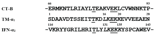
More than two decades ago octapeptide LKEKKYSP corresponding to the sequence 131-138 of human interferon-α2 (IFN-α2) capable of high affinity binding to murine thymocytes [10] and human fibroblasts [11] was obtained. Binding of labeled octapeptide was competitively inhibited by unlabeled IFN-α2, thymosin-α1 (TM-α1) and cholera toxin B-subunit (CT-B). Comparison of amino acid sequences of the octapeptide and TM-α1 showed that they contain the same LKEKK fragment corresponding to the sequence 16-20 TM-α1 and 131-135 IFN-α2 (Figure 1). We suggested that this fragment may be involved in the binding of TM-α1and IFN-α2 with a common receptor and synthetic peptide LKEKK may also have the same ability.
Recently we synthesized peptide LKEKK and found that [3H] LKEKK binds with high affinity to donor blood T lymphocytes [12,13] rat intestinal epithelial cell membranes [14,15] rat IEC-6 [16] and human Caco-2 [16,17] intestinal epithelial cells, murine Raw 264.7 macrophage-like cells [18]. Treatment of cells and membranes with proteases did not affect the [3H] LKEKK binding, suggesting the non-protein nature of the peptide receptor. The binding was completely inhibited by TM-α1, IFN-α2, and cholera toxin B subunit (CT-B). Thus, using [3H] LKEKK, we demonstrated the existence of a non-protein receptor common for TM-α1, IFN-α2, and CT-B on different cell types. We suggested that this receptor could be the CT receptor, which is known to be a GM1-glanglioside [5,6]. It was found that CT-B and the peptide: LKEKK at concentrations of 10 1000 nM increased in a dose-dependent manner the nitric oxide (NO) production and the soluble guanylate cyclase (sGC) activity in IEC-6 and Caco-2 cells [16,17]. Taking into account that NO acts as a primary activator of sGC, it can be assumed that the effect of CT-B and peptide LKEKK on the target cell is realized in the following way: increase in the iNOS expression → increase in the NO production → increase in the sGC activity → increase in intracellular levels of cGMP.
The purpose of this paper was to study the effect of CT-B and peptide LKEKK on TNF-α-induced pro-inflammatory signaling and pro-inflammatory cytokine responses in a mouse model of experimental colitis.
Materials and Methods
Peptides and Proteins
Human thymosin-α1 were obtained from Immundiagnostik AG (Germany), cholera toxin B subunit was from Sigma (USA). Peptides LKEKK and KKEKL were synthesized on an Applied Biosystems Model 430A automatic synthesizer (USA) using the Boc/Bzl tactics of peptide chain elongation as described previously [19]. The peptides were purified to homogeneous state by preparative reverse-phase HPLC (Gilson chromatograph, France) on a Delta Pack C18 column, 100A (39×150 mm, mesh size 5 ïm; flow rate 10 mL/min, elution with 0.1% TFA, gradient of acetonitrile 10–40% in 30 min). The molecular masses of peptides were determined by fast atom bombardment mass spectrometric analysis (Finnigan mass spectrometer, San Jose, CA). The data of amino acid analysis (hydrolysis by 6 M HCl, 22h, 110°C; LKB 4151 Alpha Plus amino acid analyzer, Sweden) and mass spectrum analysis are presented in Table 1.
Table 1: Main characteristics of the synthesized peptides.
| Peptide | Purity, % | Amino acid analysis data | Molecular mass, D |
| LKEKK | >98 | Glu 1.09, Leu 1.00, Lys 3.27 | Glu 1.09, Leu 1.00, Lys 3.27 |
| KKEKL | >97 | Glu 1.12, Leu 1.03, Lys 3.32 | 648.6 (644.87) |
Cell Culture
Human Caco-2 intestinal epithelial cell line was kindly provided by Moscow Research Epidemiology and Microbiology Institute. Cells were maintained at 37°C in 5% CO2 in DMEM medium supplemented with 10% fetal bovine serum. All experiments were performed with cells which were in between 15 and 25 passages. Cells in exponential growth phase used for all experiments.
Cells were seeded at a density of 1×10⁵ cells/well in 24- or 48-well plates (Corning, Lowell, MA) and grown for 5–7 days. Cell monolayers were rinsed with Hank's buffered salt solution and treated with CT-B or peptides LKEKK and KKEKL in culture medium containing 5% FBS for 2 h. To induce inflammation cells were stimulated with recombinant human TNF-α (2 ng/mL, Sigma, USA).
Animals
6–8-week-old female BALB/c mice (16–20 g) (the nursery of the Branch of Institute of Bioorganic Chemistry) were group housed on a 12-h light–dark cycle and allowed unrestricted access to standard mouse chow and water. The experiments procedures followed the guidelines for the care and use of laboratory animals, and they were approved by the Institutional Ethics Committee.
Treatment of Mice with CT-B/peptides and Induction of DSS-Induced Colitis
Mice were administered CT-B or peptide LKEKK (20 mg/kg body weight in 100 μL of water) or vehicle, by oral gavage, starting on day 1 and continuing until day 14. On day 7, colitis was induced by the addition of 5% dextran sodium sulfate (DSS) (MW 36–50 kDa, MP Biomedicals, Solon, OH) to drinking water and continued until day 14. Negative control mice received water only, and positive control mice received DSS only. Mice were euthanized on day 14. Colons were removed and measured, and tissue sections were flash frozen for further analysis.
Clinical Analysis of Colitis
Mice were weighed daily, and data are expressed as mean percentage change relative to starting body weight. Mice were monitored daily for stool consistency, presence of blood in stool or bleeding and general appearance, and a clinical activity score (ranging from 0 to 7) was calculated as described by Maxwell et al. [20].
Histological Analysis of Colitis
Paraffin sections from the distal colon were stained with hematoxylin and eosin, and a histological assessment of tissue damage and inflammation was performed as described by Maxwell et al. [20].
Cytokine ELISAs
Measurement of IL-8 in Caco-2 culture supernatants was carried out as described previously [11]. To measure the concentrations of TNF-α and IL-6 in mouse colon tissues, tissues were homogenized in three volumes of ice-cold PBS containing 1 mM PMSF, 10 μg/mL aprotinin, 10 μg/mL leupeptin, and 10 μg/mL pepstatin A (Sigma-Aldrich) using a POLYTRON® PT 1200 E (Kinematica AG., Switzerland) and centrifuged at 12,000 ×g for 10 min at 4°C. The protein concentration was determined by the Lowry method [21] using bovine serum albumin as a standard. TNF-α and IL-6 ELISAs were carried out using anti-mouse IL-6 (MP5-20F3) or anti-mouse/rat TNF-α (TN3-19.12) (BD Biosciences, San Jose, CA) according to the manufacturer's instructions.
Measurement of the guanylate cyclase activity
The guanylate cyclase (sGC) activity was measured by monitoring the conversion of [α-32P]GTP to [32P]cGMP [22]; the product was isolated by precipitation with zinc carbonate and chromatography on a column of aluminum oxide[23]. The enzyme activity was expressed as the amount of cGMP produced in 10 min (in nanomoles per 1 mg protein). The protein concentration was determined by the Lowry method [21] using bovine serum albumin as a standard. To inhibit the activity of sGC, an inhibitor of the enzyme ODQ (1Hâ€[1,2,4]oxadiazolo[4,3â€α]quinoxalinâ€1â€one, an inhibitor of soluble guanylate cyclase) [24].in the concentration range of 5-100 μM was used.
Statistical Analysis
Data are expressed as means ± SEM. Student's t-test was used when comparisons were made only between the two groups. Differences were considered significant when p < 0.05.
Results
CT-B and Peptide LKEKK Reduce TNF-α-Induced Expression of Inflammatory Mediators in Caco-2 Cells
Our experiments have shown that pre-treatment of Caco-2 cells with CT-B or peptide LKEKK (the concentration range of 100-5000 µM) significantly reduced in a dose-dependent manner TNF-α-induced IL-8 secretion (Table 2). Peptide KKEKL with inverted amino acid sequence was inactive; which indicates a high specificity of the peptide LKEKK action. In addition, treatment with CT-B or peptide LKEKK significantly reduced mRNA levels of IL-8, as well as pro-inflammatory cytokines TNF-α, IL-6, and IL-1β, when compared to cells treated with TNF-α alone (Table 3). At the same time, a significant increase in the expression of the anti-inflammatory cytokine IL-10 was also observed in response to CT-B or peptide LKEKK treatment. Tested in parallel peptide KKEKL was inactive (Table 4). Thus, CT-B and peptide LKEKK decrease the production of pro-inflammatory cytokines in vitro.
Table 2: Effect of pre-treatment of Caco-2 cells with CT-B or peptides LKEKK and KKEKL on TNF-α-induced secretion of pro-inflammatory cytokine IL-8.
| Peptide/Protein (µM) | IL-8 (pg/mL ± SEM) | ||
|---|---|---|---|
| CT-B | Peptide LKEKK | Peptide KKEKL | |
| - (Control) | 844 ±72 | ||
| 10 | 796 ±66 | 824 ±73 | 834 ±69 |
| 100 | 532 ± 53* | 586 ± 57* | 857 ±73 |
| 500 | 209 ± 17* | 233 ± 19* | 849 ±86 |
| 1000 | 122 ± 10* | 146 ± 12* | 829 ±84 |
| 5000 | 118 ± 15* | 139 ± 13* | 854 ±88 |
Note: Caco-2 cells were pre-treated for 2 h with CT-B or peptides at indicated doses, followed by stimulation with 2 ng/mL TNF-α for 4 h. IL-8 concentration in supernatants was measured by ELISA. Data are presented as mean ± SEM (n = 3 independent determinations).
* Significant differences between experience and control (P < 0.05).
Table 3: Effect of pre-treatment of Caco-2 cells with CT-B and peptide LKEKK on TNF-α-induced mRNA expression of pro-inflammatory cytokines.
| Peptide/Protein (µM) | Cytokine mRNA level (experiment/control ± SEM) | |||||||
|---|---|---|---|---|---|---|---|---|
| IL-8 | TNF-α | IL-6 | IL-1β | |||||
| CT-B | LKEKK | CT-B | LKEKK | CT-B | LKEKK | CT-B | LKEKK | |
| 10 | 0.9±0.1 | 0.9±0.1 | 1.0±0.1 | 1.0±0.1 | 1.0±0.1 | 1.0±0.1 | 1.0±0.1 | 1.0±0.1 |
| 100 | 0.7±0.1* | 0.8±0.1 | 0.8±0.1 | 0.8±0.1 | 0.9±0.1 | 0.9±0.1 | 0.8±0.1 | 0.8±0.1 |
| 500 | 0.5±0.1* | 0.6±0.1* | 0.5±0.1* | 0.6±0.1* | 0.6±0.1* | 0.7±0.1* | 0.7±0.1* | 0.7±0.1* |
| 1000 | 0.4±0.1* | 0.4±0.1* | 0.4±0.1* | 0.5±0.1* | 034±0.1* | 0.4±0.1* | 0.4±0.1* | 0.5±0.1* |
| 5000 | 0.4±0.1* | 0.5±0.1* | 0.4±0.1* | 0.4±0.1* | 0.3±0.1* | 0.4±0.1* | 0.4±0.1* | 0.5±0.1* |
Note: Caco-2 cells were pre-treated for 2 h with 0.5 mM of CT-B or peptides followed by stimulation with 2 ng/mL TNF-α for 4 h. mRNA expression was measured by real-time quantitative RT-PCR using GAPDH as the reference gene, and results are expressed as mRNA level relative to control. Data are presented as mean ± SEM (n = 3 independent determinations).
* Significant differences between experience and control (P < 0.05).
Table 4: Effect of pre-treatment of Caco-2 cells with CT-B or peptides LKEKK and KKEKL on TNF-α-induced expression of anti-inflammatory cytokine IL-10.
| Peptide/Protein (µM) | IL-10 mRNA level (experiment/control ± SEM) | ||
|---|---|---|---|
| CT-B | Peptide LKEKK | Peptide KKEKL | |
| 10 | 1.2 ± 0.2 | 1.0 ± 0.1 | 1.1 ± 0.2 |
| 100 | 1.9 ± 0.2* | 1.7 ± 0.1* | 1.0 ± 0.1 |
| 500 | 2.8 ± 0.2* | 2.5 ± 0.2* | 1.1 ± 0.2 |
| 1000 | 3.6 ± 0.3* | 3.0 ± 0.2* | 1.0 ± 0.1 |
| 5000 | 3.5 ± 0.3* | 3.2 ± 0.3* | 0.9 ± 0.2 |
Note: Caco-2 cells were pre-treated for 2 h with 0.5 mM of CT-B or peptides followed by stimulation with 2 ng/mL TNF-α for 4 h. mRNA expression was measured by real-time quantitative RT-PCR using GAPDH as the reference gene, and results are expressed as mRNA level relative to control. Data are presented as mean ± SEM (n = 3 independent determinations).
* Significant differences between experience and control (P < 0.05).
CT-B and Peptide LKEKK Reduce the Severity of Inflammation in a Mouse Model of DSS-Induced colitis
To investigate whether CT-B and peptide LKEKK could prevent inflammation in vivo, a mouse model of DSS-induced colitis was used. Mice were orally administered CT-B or peptide LKEKK (5 or 20 mg/kg body weight for 14 days) and DSS was introduced into the drinking water on day 7 to induce colitis. On day 14, tissues were collected for analysis. After receiving DSS for 7 days, positive control mice displayed characteristic signs of colitis including weight loss, diarrhea and rectal bleeding when compared to untreated control mice (negative control). Mice that received CT-B or peptide LKEKK (20 mg/kg) for 14 days showed significantly less pronounced clinical signs compared to positive control mice (Table 5). Treatment with CT-B or peptide LKEKK also attenuated DSS-induced weight loss and colon shortening.
Table 5: Effect of CT-B and peptides LKEKK on severity of clinical parameters of DSS-induced colitis in mice.
| Days after DSS start | Clinical score (score mean ± SEM) | |||
|---|---|---|---|---|
| Negative control | Positive control | CT-B | Peptide LKEKK | |
| 1 | < 0.1 | 0.3 ± 0.1 | 0.2 ± 0.2 | 0.3 ± 0.1 |
| 2 | < 0.1 | 0.5 ± 0.2 | 0.2 ± 0.1 | 0.2 ± 0.2 |
| 3 | < 0.1 | 0.9 ± 0.3 | 0.3 ± 0.2 | 0.4 ± 0.2 |
| 4 | < 0.1 | 1.6 ± 0.5 | 0.7 ± 0.2* | 0.9 ± 0.3 |
| 5 | < 0.1 | 2.8 ± 0.7 | 1.1 ± 0.3* | 1.5 ± 0.4* |
| 6 | < 0.1 | 4.3 ± 1.0. | 1.6 ± 0.5*. | 1.9 ± 0.6*. |
| 7 | < 0.1 | 5.2 ± 1.2 | 2.3 ± 0.5* | 2.5 ± 0.4* |
Note: Mice were given 20 mg/kg CT-B or peptide LKEKK orally for 14 d. To induce acute colitis, 5% DSS was added to drinking water on day 7. Clinical score was determined by assessing stool consistency, presence of blood in stool or rectal bleeding. Clinical scores ranged from 0 to 7. Data is presented as mean score ± SEM (n = 12 per group).
* Significant differences between experience and positive control (P < 0.05).
CT-B and Peptide LKEKK Reduce Secretion of DSS-induced Pro-inflammatory Mediators in Mice
Experiments have shown that both CT-B and peptide LKEKK (5 or 20 mg/kg body weight orally for 14 days) significantly reduced TNF-α and IL-6 production when compared to positive control mice (Table 6). These results are consistent with the in vitro results presented above and demonstrate the ability of the protein and peptide to have an anti-inflammatory effect in vivo.
Table 6: Effect of pre-treatment of mice with CT-B or peptide LKEKK on secretion of DSS-induced pro-inflammatory mediators.
| Peptide/Protein (mg/kg) | TNF-α (pg/mg protein ± SEM) | IL-6 (pg/mg protein ± SEM) | ||
|---|---|---|---|---|
| CT-B | Peptide LKEKK | CT-B | Peptide LKEKK | |
| Positive control | 22.3 ± 2.0 | 13.5 ± 1.2 | ||
| 5 | 17.2 ± 1.6 | 10.3 ± 0.7 | ||
| 20 | 5.5 ± 0.3* | 3.4 ± 0.3* | ||
Note: concentration of TNF-α and IL-6 in colon homogenates were measured by ELISA. Results are expressed as cytokine amount per total protein concentration. Data are presented as mean ± SEM (n=6 per group).
* P < 0.05 versus positive control.
An Inhibitor of sGC ODQ Abolishes the Inhibitory Effect of CT-B and Peptide LKEKK on TNF-α-Induced IL-8 Secretion in Caco-2 Cells
The data presented in the Table 7 show that ODQ, an inhibitor of soluble guanylate cyclase [24], suppressed in a dose-dependent manner the sGC activity as well as the inhibitory effect presence of 1 nM CT-B or LKEKK on the IL-8 secretion in the in TNF-α-stimulated Caco-2 cells. Thus, inhibition of the sGC activity leads to a loss of both CT-B and LKEKK ability to inhibit the IL-8 secretion.
Table 7: Effects of ODQ on the sGC activity and the IL-8 secretion in the presence of 1 nM CT-B or LKEKK in TNF-α-stimulated Caco-2 cells.
| ODQ (µM) | Guanylate cyclase activity (nmoles of cGMP per 1 mg protein in 10 min± SEM) | IL-8 (pg/mL ± SEM) | |
|---|---|---|---|
| CT-B (1 nM) | LKEKK (1 nM) | ||
| - | 2.4 ± 0.3 | 130 ± 12 | 139 ± 13 |
| 1 | 1.8 ± 0.2* | 155 ± 14 | 167 ± 12 |
| 3 | 0.9 ± 0.2* | 462 ± 39* | 439 ± 43* |
| 5 | 0.4 ± 0.1* | 775 ± 76* | 759 ± 73* |
| 10 | 0.3 ± 0.1* | 830 ±72* | 822 ±85* |
| 20 | 0.3 ± 0.1* | 837 ±83* | 845 ±66* |
Note: Caco-2 cells were stimulated with 2 ng/mL TNF-α for 4 h.
* Significant differences between experience and control (P < 0.05).
Discussion
There is good evidence that CT-B in animal models, as well as in clinical trials, to be effective in decreasing inflammation in inflammatory bowel disease (Crohn’s disease and ulcerative colitis). Boirivant et al. showed that oral administration of rCT-B protected against intestinal inflammation induced by trinitrobenzene sulfonic acid (TNBS), which is a mouse model of Crohn’s disease [25]. The protection seen in the TNBS colitis model was confirmed in a human clinical trial, in which rCT-B significantly decreased inflammation in mild to moderately active Crohn’s disease [26]. Thus, CT-B reduces the intestinal inflammation in vitro and in vivo, and may have significant therapeutic potential in both Crohn’s disease and ulcerative colitis.
Recently we have shown that CT-B and synthetic peptide LKEKK that corresponds to residues 16-20 in TM-α1 and 131-135 in IFN-α2 bind with high affinity and specificity to rat IEC-6 and human Caco-2 intestinal epithelial cells. The CT-B and peptide LKEKK binding to the cells leads to an increase in the sGC activity [17]. In this context, it is interesting comparative study of anti-inflammatory action of protein and peptide in vitro and in vivo, as well as clarifying the role of the sGC-dependent signal transduction pathway in the implementation of this action.
In this study we examined the potential anti-inflammatory properties of CT-B and peptide LKEKK using an in vitro model of TNF-α-induced inflammation in human Caco-2 intestinal epithelial cell line. For this, cells were treated with CT-B or peptide LKEKK at the concentration range of 10-5000 µM, and TNF-α (2 ng/mL) was added to induce inflammation. As it is known, stimulation of Caco-2 cells with TNF-α increases the expression of inflammatory mediators, including IL-8 [27-29], which was used here as a marker of inflammation. Experiments have shown that pre-treatment of Caco-2 cells with CT-B or peptide LKEKK at the concentration of 100-5000 µM significantly reduced TNF-α-induced IL-8 secretion in a dose-dependent manner (Table 2). Peptide KKEKL with inverted amino acid sequence was inactive; which indicates a high specificity of the peptide LKEKK action. Similarly, treatment with CT-B or peptide LKEKK significantly reduced mRNA levels of IL-8, as well as pro-inflammatory cytokines TNF-α, IL-6, and IL-1β, when compared to cells treated with TNF-α alone (Table 3). A significant increase in the expression of the anti-inflammatory cytokine IL-10 was also observed in response to CT-B or peptide LKEKK treatment (Table 4). Thus, the results indicate that both CT-B and peptide LKEKK can exert an anti-inflammatory effect in vitro.
We also studied the effect of CT-B and peptide LKEKK administration on DSS-induced inflammatory cytokines in the colon. Concentrations of TNF-α and IL-6 in colon tissue homogenates were measured by ELISA. Both protein and peptide (20 mg/kg body weight orally for 14 days) significantly reduced TNF-α production when compared to positive control mice (Table 6). At the same time, the mice in these groups showed less pronounced clinical signs of inflammation (Table 5). These findings are in line with our in vitro results, and demonstrate that both CT-B and peptide LKEKK can exert potent anti-inflammatory effects in vivo.
We recently showed that CT-B and peptide LKEKK increase in the sGC activity of target cells and suggested that this enzyme may be involved in the realization of their effects. To test this assumption, we investigated the effect of CT-B and peptide LKEKK on TNF-α-induced IL-8 secretion in Caco-2 cells under partial or total absence of the sGC activity. Inhibition the enzyme activity is achieved using an inhibitor of sGC ODQ which oxidizes the haem prosthetic group to which NO binds [24]. It was found that the decrease in enzyme activity was accompanied by a loss of the ability of the protein and peptide to inhibit cytokine secretion. Thus, CT-B and peptide LKEKK reduce the pro-inflammatory cytokine secretion in TNF-α-stimulated Caco-2 cells via sGC.
Declaration of interest
There is no conflict of interest that could be perceived as prejudicing the impartiality of the research reported.
Funding
This study was funded by Fundamental Research Program of the Presidium of RAS "Molecular and Cell Biology" (Grant # 0101-2014-0086).
References
- Sánchez J, Holmgren J (2008) Cholera toxin structure, gene regulation and pathophysiological and immunological aspects. Cell Mol Life Sci 65: 1347-1360. [crossref]
- Sanchez J, Holmgren J (2011) Cholera toxin - a foe & a friend. Indian J Med Res 133: 153-163. [crossref]
- Lai CY (1977) Determination of the primary structure of cholera toxin B subunit. J Biol Chem 252: 7249-7256. [crossref]
- Chester MA (1998) IUPAC-IUB Joint Commission on Biochemical Nomenclature (JCBN). Nomenclature of glycolipids--recommendations 1997. Eur J Biochem 257: 293-298. [crossref]
- Cuatrecasas P (1973) Gangliosides and membrane receptors for cholera toxin. Biochemistry 12: 3558-3566. [crossref]
- Holmgren J, Lönnroth I, Svennerholm L (1973) Tissue receptor for cholera EXOTOXIN: postulated structure from studies with G(M1) ganglioside and related glycolipids. Infect. Immun. 8: 208-214.
- Smits HH, Gloudemans AK, van Nimwegen M, Willart MA, Soullie T, et al. (2009) Cholera toxin B suppresses allergic inflammation through induction of secretory IgA. Mucosal Immunol. 2: 331-339.
- Sun JB, Czerkinsky C, Holmgren J (2010) Mucosally induced immunological tolerance, regulatory T cells and the adjuvant effect by cholera toxin B subunit. Scand. J. Immunol. 71: 1-11.
- Stratmann T (2015) Cholera toxin subunit B as adjuvant––an accelerator in protective immunity and a break in autoimmunity. Vaccines (Basel) 3: 579-596.
- Zav’yalov VP, Navolotskaya EV, Abramov VV, Galaktionov VG, Isaev IS, et al. (1991) The octapeptide corresponding to the region of the highest homology between a-interferon and thymosin-a1 effectively competes with both cytokines for common high-affinity receptors on murine thymocytes. FEBS Lett. 278: 187-189.
- Katayama S, Mine Y (2007) Antioxidative activity of amino acids on tissue oxidative stress in human intestinal epithelial cell model. J. Agric. Food Chem. 17: 8458-8464.
- Navolotskaya EV, Zinchenko DV, Zolotarev YA, Kolobov AA, Lipkin VM (2016) Binding of Synthetic LKEKK Peptide to Human T-Lymphocytes. Biochemistry (Moscow) 81: 871-875.
- Navolotskaya EV, Sadovnikov VB, Zinchenko DV, Lipkin VM, Zav'yalov VP (2017) Interaction of cholera toxin B subunit with T and B lymphocytes. Int Immunopharmacol 50: 279-282. [crossref]
- Navolotskaya EV, Sadovnikov VB, Zinchenko DV, Vladimirov VI, Zolotarev YA, et al. (2016) The LKEKK Synthetic Peptide as a Ligand of Rat Intestinal Epithelial Cell Membranes. Russ. J. Bioorg. Chem 42: 479-483.
- Navolotskaya EV, Sadovnikov VB, Zinchenko DV, Vladimirov VI, Zolotarev YA, et al. (2017) a1-Thymosin, a2-interferon, and the LKEKK synthetic peptide inhibit the binding of the B subunit of the cholera toxin to intestinal epithelial cell membranes. Russ. J. Bioorg. Chem. 43: 673-677.
- Navolotskaya EV, Sadovnikov VB, Zinchenko DV, Vladimirov VI, Zolotarev YA, et al. (2018) Interaction of Cholera Toxin B Subunit with Rat Intestinal Epithelial Cells. Russ. J. Bioorg. Chem. 44: 403-407.
- Navolotskaya EV, Sadovnikov VB, Lipkin VM, Zav'yalov VP (2018) Binding of cholera toxin B subunit to intestinal epithelial cells. Toxicology in Vitro 47: 269-273.
- Navolotskaya EV, Sadovnikov VB, Zinchenko DV, Vladimirov VI, Zolotarev YA, et al. (2019) Action of the B Subunit of the Cholera Toxin on the Raw 264.7 Murine Macrophage-Like Cell Line. Rus. J. Bioorg. Chem. 45: 122–128.
- Schnolzer M, Alewood P, Jones A, Alewood D, Kent SB, et al. (1992) In situ neutralization in Boc-chemistry solid phase peptide synthesis. Rapid, high yield assembly of difficult sequences. Int. J. Peptide Protein Res. 40: 180-193.
- Maxwell JR, Brown W, Smith CL, Byrne FR, Viney JL, et al. (2009) Methods of inducing inflammatory bowel disease in mice. Curr. Protoc. Pharmacol. S47: 5.58.1-5.58.37.
- LOWRY OH, ROSEBROUGH NJ, FARR AL, RANDALL RJ (1951) Protein measurement with the Folin phenol reagent. J Biol Chem 193: 265-275. [crossref]
- Schultz G, B?hme E (1984) Methods of Enzymatic Analysis. Verlag Chemie, Weinheim, Germany, pp. 379-389.
- Southam E (2001) Measurement of cGMP and soluble guanylyl cyclase activity. Curr. Protoc. Toxicol. 10: 10.5.
- Feelisch M, Kotsonis P, Siebe J, Clement B, Schmidt HH, et al. (1999) The Soluble Guanylyl Cyclase Inhibitor 1H-[1,2,4]oxadiazolo[4,3,-a]quinoxalin-1-one Is a Nonselective Heme Protein Inhibitor of Nitric Oxide Synthase and Other Cytochrome P-450 Enzymes Involved in Nitric Oxide Donor Bioactivation. Mol. Pharmacol. 56: 243-253.
- Boirivant M, Fuss IJ, Ferroni L, de Pascale M, Strober W, et al. (2001) Oral administration of recombinant cholera toxin subunit B inhibits IL-12-mediated murine experimental (trinitrobenzene sulfonic acid) colitis. J. Immunol. 166: 3522-3532.
- Stal P, Befrits R, Ronnblom A, Danielsson A, Suhr O, et al. (2010) Clinical trial: The safety and short-term efficacy of recombinant cholera toxin B subunit in the treatment of active Crohn’s disease. Aliment. Pharmacol. Ther. 31: 387-395.
- Treede I, Braun A, Jeliaskova P, Giese T, Füllekrug J, et al. (2009) TNF-a-induced up-regulation of pro-inflammatory cytokines is reduced by phosphatidylcholine in intestinal epithelial cells. BMC Gastroenterol. 9: 53.
- Baldauf KJ, Royal JM, Hamorsky KT, Matoba N (2015) Cholera toxin B: one subunit with many pharmaceutical applications. Toxins 7: 974-996.
- Zav’yalov VP, Navolotskaya EV, Vasilenko RN, Abramov VM, Volodina EY, et al. (1995) The sequence 130-137 of human interferon-a2 is involved in the competition of interferon, prothymosin a and cholera toxin B subunit for common receptors on human fibroblasts. Mol. Immunol. 32: 425-431.