Pruritus, A Frequent Symptom
Fabiola Ramallo Jadue1*, David El-Qutob López2
1 Specialist in Allergy, Immunopathology and Dermatology, U.M.R.P.S.F.X.CH, BOLIVIA.
2 Unit of Allergy – Service of Internal Medicine, Hospital de la Plana, Vila-Real (Castellón), Spain.
*Corresponding Author: Fabiola Ramallo Jadue, Professor & Specialist in Allergy, Immunopathology and Dermatology, U.M.R.P.S.F.X.CH, BOLIVIA, TEL: +591 72875286 ; FAX: +591 72875286;E-mail:fabiolaramallo3@gmail.com
Citation: Fabiola Ramallo Jadue, David El-Qutob López (2018) Pruritus, A Frequent Symptom. Allergy drugs clin immunol 2:110.
Copyright:© 2018 Fabiola Ramallo Jadue, et al. This is an open-access article distributed under the terms of the Creative Commons Attribution License, which permits unrestricted use, distribution, and reproduction in any medium, provided the original author and source are credited.
Received date: August 03, 2018; Accepted date: August 13, 2018; Published date: August 16, 2018
Abstract
The sensation of itching or itching depends still little-known mechanisms. It was considered a minor form of pain related to alterations of the cutaneous vasomotilidad. However, many reasons would indicate that it is an autonomous sensation, since pruritus and pain can coexist in the same area. Pruritus may upset the sufferer; The lower the patient, the more intense the suffering.
Keywords
Pruritus. Pain. Inflammatory mediators
Introduction
Itching is one of the most frequent manifestations of both non-dermatological and dermatological disorders, however little is even what is known about their mechanisms of production and therefore of specific forms of relief.
The itching has been historically defined as feeling that leads to the desire to scratch. It is probably the most common symptom of skin disorders but even so it has been widely ignored in the scientific research, perhaps since despite the passage of time, still satisfactory explanations not found about their production tooling.
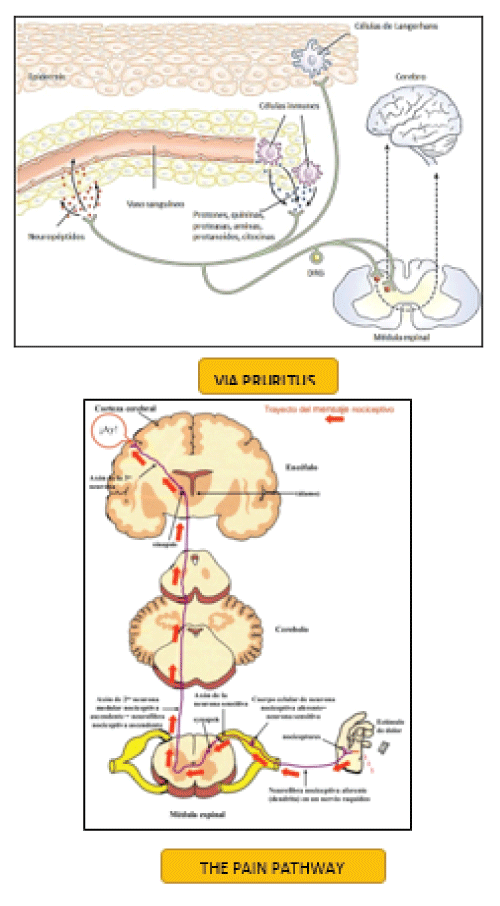
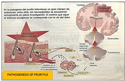
Many consider the itching as a pain-related symptom, and both apparently share a common nerve pathway [1]. However, it is difficult to explain then why morphine suppresses pain but increases or even induce itching. It has been suggested that the effect prurigeno of morphine is consequence of the release of histamine from mast cells, although pruritus induced by this drug not is relieved by the administration of antihistamines. Figure 1.
The truth is that it is still much to elucidate as pruritus and shortage of convincing animal models makes this symptom remains largely a mystery.
Spontaneous itching, itchy skin, and aloquinesis
The application of a stimulus capable of causing itching resulting in two kinds of clearly defined responses: on the one hand an itching well located at the site of the stimulus that lingers briefly after the stimulus stopped and, secondly, an itchy area diffuse, little defined, that surrounds the stimulated site and not itching spontaneously but soft touch and the friction [2]. Brickford, in 1938, appointed to these two areas as spontaneous pruritus and itchy skin, respectively. LaMotte, in 1992, introduced the term aloquinesis to refer to the area of itchy skin, in parallel with the term allodynia, which refers to the feeling of pain caused by mild rubbing of the skin.
The aloquinesis has been explained as the result of the excitation of Central neurons mediating of pruritus through receiving Interneurons of the stimulus from of fast driving and low-threshold mechanoreceptors, which would respond to the histamine acting about skin-free terminations.
The phenomenon of spontaneous pruritus and itchy skin or aloquinesis is an element common, present in many dermatological conditions [3]. The clearest example is the insect bite, in where you can see a hive with spontaneous pruritus defined surrounded by a more extensive diffuse area of aloquinesis that persists for hours or even days after you Welt it has given.
Probable mediators involved in itching
In many cases this symptom is related to the inflamed skin, which led to investigate mediators of inflammation were capable of causing itching. It deepened the knowledge about the inflammation, the list of candidates potentially prurigenos was increased.
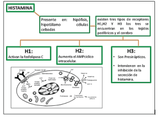
Histamine was probably involved in the physiopathogeny of pruritus, starting in 1927, Lewis discovered superficial injection of this substance on the skin caused first. Two histamine receptors in the skin (H1 and H2) is known to be involved only in pruritus type H1 [4]. Other receivers (H3 Hic) would not be represented at the skin surface, and therefore would have no influence on the development of the itching. Figure 2.
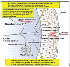
The main sources of histamine are mast cells, cells that synthesize and store this substance in their cytoplasmic grains [5]. The release of histamine from mast cells is the result of the union of the immunoglobulin E its (Fc) receptors on the membrane of these cells, either through the complement (C5a) or some taquininas, including substance P. Figure 3.
A wide variety of pruritic dermatoses, with inflamed skin as urticaria retrieves large amounts of histamine, and therein has been shown symptomatic relief before the administration of H1 antagonists.
Prostaglandin E2 (PGE2) is located in high concentrations in many inflammatory dermatoses attending itchy as psoriasis, contact dermatitis, and skin irradiated with ultraviolet B [6]. However, the mechanism by which the PGE2 cause this symptom would be indirect, lowering the threshold of the skin to the sensation of itching caused by histamine and other mediators. Figure 4.
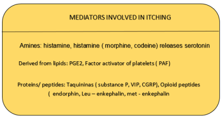
The included taquininas related to the gene for calcitonin (CGRP) and vasoactive intestinal peptide (VIP), peptide, substance P are located in nociceptive neurons-free nerve endings not myelinated in the surface of the skin. These free endings are capable of producing pain and itching [7]. The CGRP and VIP are unable per will cause itching, although both can cause inflammation. There is some evidence that the substance P, a peptide of 11 amino acids, is involved in the genesis of pruritus in certain circumstances. The intradermal injection of this substance leads to the formation of a hive erythematous and itchy although probably this occurs, at least in part, through the release of histamine by mast cells [8]. The action of substance P on these would be independent of the IGE-dependent mechanism. Figure 5.
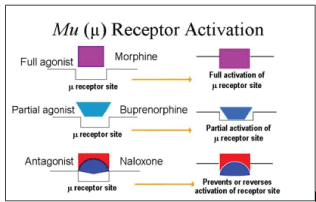
Opioid peptides are undoubtedly involved in the physiopathogeny of pain and several researchers believe that they would also intervene in the itching.
These opioids Act through the interaction of their receptors or at the level of the central nervous system, but also would have some influence at the peripheral level to enhance the itching caused by other mediators [9]. Histamine-induced itch can worsen if previously applied on the skin a low concentration of opioid peptides; This effect is antagonized by naloxone and is not mediated by receptors U. Figure 6.
Histamine, substance P, PGE2 and opioid peptides are probably the strongest candidates to be designated as mediators of the inflamed skin itchy [10]. However, there are a number of conditions in which pruritus not associated with inflammation. Such is the case of itching of the elderly, aquagenic pruritus or pruritus solar. In other situations the itching is associated with systemic diseases without cutaneous inflammation such as Cholestasis, chronic renal failure, thyrotoxicosis and Polycythemia vera. Figure 7.
In the elderly the water content of the skin tends to be decreased, even without obvious xeroderma, and that may be the cause of the itching. In aquagenic pruritus, wet skin with water at any temperature it causes distressing itching immediately out of the bath or shower and lasts about an hour, without visible signs on the skin [11]. In many of these patients have found elevated levels of histamine in blood during these episodes. Pruritus and cholestasis is so intense and persistent that it tends to become the main problem for affected patients [12]. It has been shown that this symptom is associated with high levels of bile salts since it relieved by treatment with cholestyramine.
Pruritus in chronic renal failure is harder to explain, since although in the majority of cases it is relieved after dialysis it does not correlate with plasma levels of urea and creatinine.
Some authors have related pruritus with serum of parathormone levels, since these tend to be higher in those who suffer it, Although the correlation levels of parathormone - itching is bad [13]. Histologically the skin of these patients shows higher concentration of mast cells and an abnormal pattern of cutaneous nerve terminals, although the latter may be a consequence of the permanent scratching.
Pruritus in the thyrotoxicosis is often widespread and intractable. In many cases it relates to the increase of the cutaneous blood flow which in turn increases the skin temperature, reducing the threshold for pruritus [14].
Itching of Polycythemia vera affects about half of patients with this disease, appears in the form characteristic after the bath with water at any temperature and lasts between half an hour and an hour [15]. Visible changes in the skin are not appreciated and commonly found elevated levels of histamine during these episodes.
Central and peripheral nervous mechanisms involved in itching
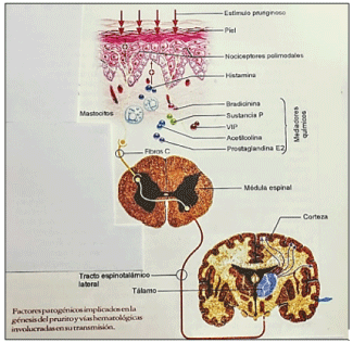
While many have linked to the nerve pathways of pain and itch it seems that fibres C nociceptive polimodal different in both cases would be involved. A subset of these fibers that would transmit only pain and other equivalent subgroup that transmit sensations of itching would be. Primary neurons make synapses with secondary neurons in the dorsal horns of the spinal cord, from there it would connect with the contralateral thalamus through the espinotalamicolateral tract [16]. There are tertiary neurons (thalamocortical tracts) that would serve as a relay for the sensations of itching on his way to the cortex.
The scrape response to pruritus, although is modulated by higher education institutions, would be rather a spinal reflex. After scratching, itching be relieved for 15 to 25 minutes, perhaps the time required for the regeneration of damaged nerve endings [17]. The itching would result from the combination of the activation of the C firas and the loss of central inhibition. It is likely that scratching is a specific suppressor both centrally and segmental local.
The simple explanation described above has historically accepted: a stimulus causes itching, stimulates nerve endings and is transmitted progressively toward the spinal cord and from there to the cerebral cortex. However, have not been identified so far such specific fibres for the itching [18]. On the other hand, there would be an active participation of the central nervous system in the genesis of pruritus, regardless of the peripheral stimulation, which would explain the many circumstances in which this symptom without such stimulation peripheral. The mechanism is likely to be more complex and could be summarized in the following manner: some agents would be able to stimulate itch through his peripheral action, and on the other hand in certain (probably systemic) disorders such stimulation feeling would act at the central level, on the inhibitory mechanisms of cortical, producing pruritus without the need for a stimulus at the peripheral level. Figure 8.
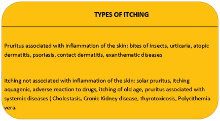
Figure 8
Treatment of pruritus
Itching treatment will always depend on the underlying cause and whether or not is associated with an inflammation of the skin.
Overall itching is usually relieved decreasing the temperature of the skin, through the use of light clothing, one year before going to sleep, adequate ventilation of the environments and the application of creams or lotions refreshing.
The efficacy of theoretically anti-itch topical such as menthol or phenol substances is unknown [19]. And some authors consider that acupuncture or Transcutaneous electrical stimulation may be helpful in intractable cases of pruritus.
The pruritic dermatoses most commonly used antihistamines, but there is still controversy about the appropriateness of using anti H1, sedatives or not sedatives. In situations in which pruritus is predominantly nocturnal, or this determines when feelings of nervousness and anxiety, they tend to be most useful sedative antihistamines [20].
The use of topical corticosteroids is reserved for those patients with pruritus associated with Dermatologic inflammatory disorders. Systemic corticosteroids low power in association with antihistamines tend to be of help in acute periods of skin inflammation.
In certain cases of intractable pruritus by disorders such as amyloidosis, cutaneous or in some Lymphoma patients with psoriasis and atopic dermatitis have been employed successfully opioid antagonists such as naltrexone.
Conclusions
The pruritus can be a symptom of numerous general affections (as uremia, diabetes, colestasis, leucosis, mieloma, Hodking's disease, chronic poisonings) and of changed dermatosis (allergic, autoimmune, parasitic, different), or to represent a disease in if same. The first aim consists of specifying the reason for: to eliminate it (as the parasitosis), to neutralize it (as in case of controlling the effects of a chemical mediator) and controlling his result (acting in form adapted on the effects of the plastered one), to break the vicious circle of pruritus - scratched - cutaneous injury - pruritus).
References
- Van Loey NEE, Bremer M, Faber AW, Middelkoop E, Nieuwenhuis MK, et al. (2008) Itching following burns: epidemiology and predictors. Br J Dermatol 158: 95-100.
- Willebrand M, Low A, Dyster-Aas J, Kildal M, Andersson G, et al. (2004) Pruritus, personality traits and coping in longterm follow-up of burn injured patients. Acta Derm Venereol 84: 375-380.
- Bell L, McAdams T, Morgan R, Parshley PF, Pike RC, et al. (1988) Pruritus in burns: a descriptive study. J Burn Care Rehabil 9: 305-308. [crossref]
- Jung SI, Seo CH, Jang K, Ham BJ, Choi IG, et al. (2009) Efficacy of naltrexone in the treatment of chronic refractory itching in burn patients: preliminary report of an open trial. J Burn Care Res 30: 257-260.
- Goutos I (2009) Re: 'Scratching the surface-managing the itch associated with burns: a review of current knowledge' [Burns 34 (2008) 751-760]. Burns 35: 754-755. [crossref]
- Malenfant A, Forget R, Papillon J, Amsel R, Frigon JY, et al. (1996) Prevalence and characteristics of chronic sensory problems in burn patients. Pain 67: 493-500. [crossref]
- Dalgard F, Svensson A, Holm JO, Sundby J (2004) Self-reported skin morbidity among adults: associations with quality of life and general health in a Norwegian survey. J Investig Dermatol Symp Proc 9: 120-125.
- Schmelz M, Schmidt R, Weidner C, Hilliges M, Torebjork HE, et al. (2003) Chemical response pattern of different classes of C-nociceptors to pruritogens and algogens. J Neurophysiol 89: 2441-2448. [crossref]
- Ikoma A, Steinhoff M, Ständer S, Yosipovitch G, Schmelz M (2006) The neurobiology of itch. Nat Rev Neurosci 7: 535-547. [crossref]
- Toyoda M, Nakamura M, Makino T, Hino T, Kagoura M, et al. (2002) Nerve growth factor and substance P are useful plasma markers of disease activity in atopic dermatitis. Br J Dermatol 147: 71-79. [crossref]
- Oaklander AL, Bowsher D, Galer B, Haanpää M, Jensen MP (2003) Herpes zoster itch: preliminary epidemiologic data. J Pain 4: 338-343. [crossref]
- Revista El Dolor - Nº 57 - Año 21 - Julio 2012
- Rees JL, Laidlaw A (1999) Pruritus: more scratch than itch. Clin Exp Dermatol 24: 490-493. [crossref]
- Sassmannshausen J, Lim HW (1999) Action spectrum and response to antihistamine use in solar pruritus. Arch Dermatol 135: 1122-1123. [crossref]
- Tang WY, Chan LY, Lo KK, Wong TW (1999) Evaluation on the antipruritic role of transcutaneous electrical nerve stimulation in the treatment of pruritic dermatoses. Dermatology 199: 237-241. [crossref]
- Metze D, Reimann S, Beissert S, Luger T (1999) Efficacy and safety of naltrexone, an oral opiate receptor antagonist, in the treatment of pruritus in internal and dermatological diseases. J Am Acad Dermatol 41: 533-539. [crossref]
- Fabrizi G, Romano A, Vultaggio P, Bellegrandi S, Paganelli R, et al. (1999) Heterogeneity of atopic dermatitis defined by the immune response to inhalant and food allergens. Eur J Dermatol 9: 380-384. [crossref]
- Jappe U, Heuck D, Witte W, Gollnick H (1998) Superantigen production by Staphylococcus aureus in atopic dermatitis: no more than a coincidence? J Invest Dermatol 110: 844-846. [crossref]
- Mattila JO, Vornanen M, Vaara J, Katila ML (1996) Mycobacteria in prurigo nodularis: the cause or a consequence? J Am Acad Dermatol 34: 224-228. [crossref]
- Tanaka M, Aiba S, Matsumura N, Aoyama H, Tagami H (1995) Prurigo nodularis consists of two distinct forms: early-onset atopic and late-onset non-atopic. Dermatology 190: 269-276. [crossref]