Design of Novel Cancer Inhibitors Using Molecular Docking, Dynamics Simulation and 3D-QSAR and 3D-QSPR Studies
Alireza Heidari1,2,3,4*, Zahra Torfeh5
1Faculty of Chemistry, California South University, 14731 Comet St. Irvine, CA 92604, USA.
2BioSpectroscopy Core Research Laboratory (BCRL), California South University, 14731 Comet St. Irvine, CA 92604, USA.
3Cancer Research Institute (CRI), California South University, 14731 Comet St. Irvine, CA 92604, USA.
4American International Standards Institute (AISI), Irvine, CA 3800, USA.
5An Independent, Volunteer and Unaffiliated Researcher.
*Corresponding Author: Alireza Heidari, Faculty of Chemistry, California South University, 14731 Comet St. Irvine, CA 92604, USA, BioSpectroscopy Core Research Laboratory (BCRL), California South University, 14731 Comet St. Irvine, CA 92604, USA, Cancer Research Institute (CRI), California South University, 14731 Comet St. Irvine, CA 92604, USA, American International Standards Institute (AISI), Irvine, CA 3800, USA,Tel: +1 408-816-2779; Fax: 00-40-253-210 +1 408-816-2779 432; E-mail: scholar.researcher.scientist@gmail.com; alireza.heidari@calsu.us; central@aisi-usa.org
Citation: Alireza Heidari, Zahra Torfeh (2023) Design of Novel Cancer Inhibitors Using Molecular Docking, Dynamics Simulation and 3D-QSAR and 3D-QSPR Studies. Arch Mol Med & Gen 5: 113.
Received: January12, 2023; Accepted: January 20, 2023; Published: January 23, 2023.
Copyright: © 2023 Alireza Heidari, et al. This is an open-access article distributed under the terms of the Creative Commons Attribution License, which permits unrestricted use, distribution, and reproduction in any medium, provided the original author and source are credited.
Graphical Abstract
Considering the importance of oxadiazole derivatives as effective anti–cancer Nano drugs on cancer cells and various other therapeutic effects, in this research, the effect of new oxadiazole derivatives called 3–(4–chlorophenyl)–5–(4–fluorophenyl)–4–phenyl–4,5–dihydro–1,2,4–oxadiazole and 3,5–bis–(4–chlorophenyl)–4–phenyl–4,5–dihydro–1,2,4–oxadiazole on single–stranded DNA/RNA in a solution. We studied the use of different spectroscopic methods. The present study investigated the effect of 3–(4–chlorophenyl)–5–(4–fluorophenyl)–4–phenyl–4,5–dihydro–1,2,4–oxadiazole and 3,5–bis–(4–chlorophenyl)–4–phenyl–4,5–dihydro–1,2,4–oxadiazole on single–stranded DNA/RNA in laboratory conditions. The results show that the absorption rate of single–stranded DNA/RNA increases due to the interaction with 3–(4–chlorophenyl)–5–(4–fluorophenyl)–4–phenyl–4,5–dihydro–1,2,4–oxadiazole and 3,5–bis–(4–chlorophenyl)–4–phenyl–4,5–dihydro–1,2,4–oxadiazole at 210 and 260 (nm) wavelengths. The emission spectrum of single–stranded DNA/RNA increases in a concentration–dependent trend of 3–(4–chlorophenyl)–5–(4–fluorophenyl)–4–phenyl–4,5–dihydro–1,2,4–oxadiazole and 3,5–bis–(4–chlorophenyl)–4–phenyl–4,5–dihydro–1,2,4–oxadiazole, which indicates the binding of 3–(4–chlorophenyl)–5–(4–fluorophenyl)–4–phenyl–4,5–dihydro–1,2,4–oxadiazole and 3,5–bis–(4–chlorophenyl)–4–phenyl–4,5–dihydro–1,2,4–oxadiazole with chromophores present in single–stranded DNA/RNA. The present study investigated the effect of 3–(4–chlorophenyl)–5–(4–fluorophenyl)–4–phenyl–4, 5–dihydro–1,2,4–oxadiazole and 3,5–bis–(4–chlorophenyl)–4–phenyl–4,5–dihydro–1,2,4–oxadiazole on single–stranded DNA/RNA in laboratory conditions. The results show that the absorption rate of single–stranded DNA/RNA increases due to the interaction with 3–(4–chlorophenyl)–5–(4–fluorophenyl)–4–phenyl–4,5–dihydro–1,2,4–oxadiazole and 3,5–bis–(4–chlorophenyl)–4–phenyl–4,5–dihydro–1,2,4–oxadiazole at 210 and 260 (nm) wavelengths. The emission spectrum of single–stranded DNA/RNA increases in a process dependent on the concentration of 3–(4–chlorophenyl)–5–(4–fluorophenyl)–4–phenyl–4,5–dihydro–1,2,4–oxadiazole and 3,5–bis–(4–chlorophenyl)–4–phenyl–4,5–dihydro–1,2,4–oxadiazole, which indicates the binding of 3–(4–chlorophenyl)–5–(4–fluorophenyl)–4–phenyl–4,5–dihydro–1,2,4–oxadiazole and 3,5–bis–(4–chlorophenyl)–4–phenyl–4,5–dihydro–1,2,4–oxadiazole with chromophores present in single–stranded DNA/RNA. The results obtained from the effect of 3–(4–chlorophenyl)–5–(4–fluorophenyl)–4–phenyl–4,5–dihydro–1,2,4–oxadiazole and 3,5–bis–(4–chlorophenyl)–4–phenyl–4,5–dihydro–1,2,4–oxadiazole on single–stranded DNA/RNA can provide useful information in the field of designing anti–cancer Nano drugs with oxadiazole derivatives with more anti–tumor effect and less side effects.
Figure: 1
Figure: 2
Figure: 3
Schematic of the 3–(4–chlorophenyl)–5–(4–fluorophenyl)–4–phenyl–4,5–dihydro–1,2,4–oxadiazole and 3,5–bis–(4–chlorophenyl)–4–phenyl–4,5–dihydro–1,2,4–oxadiazole as anti–cancer Nano drugs’ effect and delivery mechanism on DNA/RNA in human breast cancer cells.
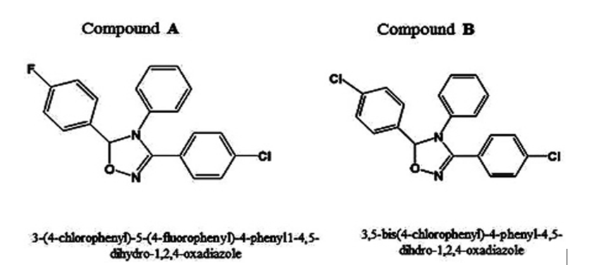
Molecular structures of (a) 3–(4–chlorophenyl)–5–(4–fluorophenyl)–4–phenyl–4,5–dihydro–1,2,4–oxadiazole and (b) 3,5–bis–(4–chlorophenyl)–4–phenyl–4,5–dihydro–1,2,4–oxadiazole.
Keywords
Molecular Dynamics, Simulation, Perception, Binding Affinity, Performance, Nano Synthesized, DNA/RNA, Human Cancer Cells, Biospectroscopic Methods and Techniques.
Introduction
Despite the efforts of scientists and the advancement of science and technology, cancer is still one of the deadliest diseases of mankind, and therefore, research on this disease and its treatment methods are of great interest. Therefore, it can be important to identify the mechanism of action of anti–cancer Nano drugs in order to use them more effectively in the treatment of various types of cancer. The nucleus is the most important organelle that exists in eukaryotic cells, and the target of many new compounds as well as anti–cancer Nano drugs is the cell nucleus. DNA/RNA inside the nucleus of eukaryotic cells during replication or transcription is bare [1–38]. Normally, it is accompanied by histone and non–histone proteins, which together form structures called nucleosomes, which are called chromatin. Therefore, anti–tumor Nano drugs can target either single–stranded or double–stranded DNA/RNA to perform their action. Oxadiazoles are widely found in biology, medicine and polymer science. The most well–known and important oxadiazole are 3–(4–chlorophenyl)–5–(4–fluorophenyl)–4–phenyl–4,5–dihydro–1,2,4–oxadiazole and 3,5–bis–(4–chlorophenyl)–4–phenyl–4,5–dihydro–1,2,4–oxadiazole, which is prescribed in low doses as a blood thinner. Several oxadiazole are used as anti–cancer Nano drugs in modern and recent medicine. Oxadiazole and its derivatives also show a wide range of different physiological activities, including anti–inflammatory, anti–bacterial, anti–cancer, anti–clotting and anti–HIV activities. Oxadiazole and its derivatives also have a wide range of activities. They show various physiological properties such as anti–inflammatory, anti–bacterial, anti–cancer, anti–clotting, anti–HIV, anti–viral, anti–viral activities [39–57]. They are also used as ingredients in perfumes, cosmetics, food additives, pharmaceuticals, in the preparation of insecticides, optical brighteners, and diffused and laser fluorescent dyes. become oxadiazoles have been noticed due to their toxicity and carcinogenicity. In addition, they show photodynamic effects and are useful intermediates for the synthesis of 3–(4–chlorophenyl)–5–(4–fluorophenyl)–4–phenyl–4,5–dihydro–1,2,4–oxadiazole and 3,5–bis–(4–chlorophenyl)–4–phenyl–4,5–dihydro–1,2,4–oxadiazole. Tetrazoles participate in the pharmacokinetics and metabolism of anti–cancer Nano drug delivery [58–76]. Therefore, they are always a good candidate for binding and acting on protein and DNA/RNA. 3–(4–chlorophenyl)–5–(4–fluorophenyl)–4–phenyl–4,5–dihydro–1,2,4–oxadiazole and 3,5–bis–(4–chlorophenyl)–4–phenyl–4,5–dihydro–1,2,4–oxadiazole are oxadiazole derivatives successfully synthesized via domino Knoevenagel. These oxadiazole derivatives participate in the formation of free radicals in skin cells and causes damage to the structure of DNA/RNA and protein in the cell, which then causes cancer in humans. Considering the above, the purpose of this experiment is to investigate the effect of these oxadiazole derivatives on the single–stranded DNA/RNA of calf thymus gland. The use of calf thymus gland, due to the high amount of DNA/RNA compared to protein, allowed us to investigate the effect of 3–(4–chlorophenyl)–5–(4–fluorophenyl)–4–phenyl–4,5–dihydro–1,2,4–oxadiazole and 3,5–bis–(4–chlorophenyl)–4–phenyl–4,5–dihydro–1,2,4–oxadiazole on pure DNA/RNA [77–114].
Materials, Methods and Techniques
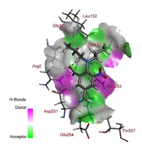
3–(4–chlorophenyl)–5–(4–fluorophenyl)–4–phenyl–4,5–dihydro–1,2,4–oxadiazole and 3,5–bis–(4–chlorophenyl)–4–phenyl–4,5–dihydro–1,2,4–oxadiazole as oxadiazole are oxadiazole derivatives that are synthesized through the reaction of active carbonylated compounds such as Isatin derivatives, aromatic aldehydes with Malononitrile through a multicomponent domino reaction. (Knoevenagel condensation/dipolar 1 and 3 ring addition) is performed without using any catalysis in water solvent at a temperature of 50 degrees Celsius. 2 mg of oxadiazole derivatives were combined with 1 ml Tris buffer solution purchased from Merck, Germany, and the solution was kept in the refrigerator. Thymus single–stranded DNA/RNA was purchased from SIGMA. To prepare the DNA/RNA solution, 2 mg of DNA/RNA powder was mixed with 1 ml of 0.01 M Tris buffer with pH 7.4 and the solution was kept in the refrigerator. The interaction of 3–(4–chlorophenyl)–5–(4–fluorophenyl)–4–phenyl–4,5–dihydro–1,2,4–oxadiazole and 3,5–bis–(4–chlorophenyl)–4–phenyl–4,5–dihydro–1,2,4–oxadiazole with single–stranded DNA/RNA was carried out using 10 mM Tris acid buffer (pH 7.2, at room temperature and away from light). For this purpose, a constant concentration of single–stranded DNA/RNA was prepared and incubated with different concentrations of 3–(4–chlorophenyl)–5–(4–fluorophenyl)–4–phenyl–4,5–dihydro 1,2,4–oxadiazole and 3,5–bis–(4–chlorophenyl)–4–phenyl–4,5–dihydro–1,2,4–oxadiazole for one hour, and after the desired time, it was used for UV–Vis spectroscopy studies. The absorbance of the solution resulting from the interaction of 3–(4–chlorophenyl)–5–(4–fluorophenyl)–4–phenyl–4,5–dihydro–1,2,4–oxadiazole and 3,5–bis–(4–chlorophenyl)–4–phenyl–4,5–dihydro–1,2,4–oxadiazole with single–stranded DNA/RNA and also the absorbance of different concentrations of the anti–cancer Nano drug in acidic Tris buffer at 210 and 260 (nm) using the spectrophotometer Carry 100 model Bio–Varian and the production of the country of Australia was read and after performing the necessary calculations, the relevant curves were drawn. In order to conduct fluorescence spectroscopy studies, first, a fixed concentration of single–stranded DNA/RNA was incubated with different concentrations of 3–(4–chlorophenyl)–5–(4–fluorophenyl)–4–phenyl–4,5–dihydro–1,2,4–oxadiazole and 3,5–bis–(4–chlorophenyl)–4–phenyl–4,5–dihydro–1,2,4–oxadiazole at room temperature and away from light for one hour. After a certain period of time has passed, the solution resulting from the interaction of 3–(4–chlorophenyl)–5–(4–fluorophenyl)–4–phenyl–4,5–dihydro–1,2,4–oxadiazole and 3,5–bis–(4–chlorophenyl)–4–phenyl–4,5–dihydro–1,2,4–oxadiazole with single–stranded DNA/RNA is excited at a wavelength of 258 (nm) and their emission spectrum is in the range of 200 to 700 (nm) using a spectrofluorometer device. Carry Eclipse, Bio–Varian model, made in Australia. The solution resulting from the interaction of single–stranded DNA/RNA with 3–(4–chlorophenyl)–5–(4–fluorophenyl)–4–phenyl–4,5–dihydro–1,2,4–oxadiazole and 3,5–bis–(4–chlorophenyl)–4–phenyl–4,5–dihydro–1,2,4–oxadiazole was also drawn at the excitation wavelength of 258 (nm) and its fluorescence emission spectrum was drawn in the range of 500–550 (nm). Also, the emission spectrum of the standard samples of 3–(4–chlorophenyl)–5–(4–fluorophenyl)–4–phenyl–4,5–dihydro–1,2,4–oxadiazole and 3,5–bis–(4–chlorophenyl)–4–phenyl–4,5–dihydro–1,2,4–oxadiazole was drawn in the mentioned range and then it was subtracted from the emission of the treated samples. During the experiments, excitation and emission slits were considered to be 10 and 5 (nm), respectively, and a quartz cuvette with a width of one centimeter was used. After drawing the curves resulting from the interaction, according to the obtained information, the Io–I/Io×100 curve was drawn against different concentrations of the anti–cancer Nano drug. In this formula, Io is the intensity of fluorescence emission in the absence of anti–cancer Nano anti–cancer Nano drug and I is the intensity of fluorescence emission in the presence of different concentrations of the anti–cancer Nano drug. Also, the constant of the Stern–Volmer equation (Ksv) was calculated to estimate the amount of fluorescence quenching. In the Stern–Volmer equation Io/I = 1+ Ksv [Q], Io and I are respectively the amount of intrinsic emission in the absence and presence of the quencher 3–(4–chlorophenyl)–5–(4–fluorophenyl)–4–phenyl–4,5–dihydro–1,2,4–oxadiazole and 3,5–bis–(4–chlorophenyl)–4–phenyl–4,5–dihydro–1,2,4–oxadiazole, [Q] the quenching concentration Ksv is the Stern–Volmer quenching constant of quenching–exposed Fluorines. Accordingly, Ksv for 3–(4–chlorophenyl)–5–(4–fluorophenyl)–4–phenyl–4,5–dihydro–1,2,4–oxadiazole and 3,5–bis–(4–chlorophenyl)–4–phenyl–4,5–dihydro–1,2,4–oxadiazole as the slope of the Io/I plot is different concentrations of 3–(4–chlorophenyl)–5–(4–fluorophenyl)–4–phenyl–4,5–dihydro–1,2,4–oxadiazole and 3,5–bis–(4–chlorophenyl)–4–phenyl–4,5–dihydro–1,2,4–oxadiazole were obtained. In order to study the second structure change, after the interaction of single–stranded DNA/RNA with 3–(4–chlorophenyl)–5–(4–fluorophenyl)–4–phenyl–4, 5–dihydro–1,2,4–oxadiazole and 3,5–bis–(4–chlorophenyl)–4–phenyl–4,5–dihydro–1,2,4–oxadiazole, CD spectroscopy was used. CD study in the Near–UV region and preferably in the wavelength range of 200–320 (nm) and using AVIV model 215 spectropolarimeter with a quartz cuvette with a width of 1 cm for the near region and a cuvette with a width of 1 mm for It was carried out in the remote area under a continuous flow of nitrogen gas and at a temperature of 23 degrees Celsius. The spectrum of interaction samples from the spectrum of standard samples of 3–(4–chlorophenyl)–5–(4–fluorophenyl)–4–phenyl–4,5–dihydro–1,2,4–oxadiazole and 3,5–bis–(4–chlorophenyl)–4–phenyl–4,5–dihydro–1,2,4–oxadiazole anti–cancer Nano drug that were drawn under the same conditions, the fraction and data under the title (Molar Ellipticity) and in the form of [?]: deg×cm2×dmol–1 were reported (Figures 1–9).
Figure1: Modelling of (a) 3–(4–chlorophenyl)–5–(4–fluorophenyl)–4–phenyl–4,5–dihydro–1,2,4–oxadiazole and (b) 3,5–bis–(4–chlorophenyl)–4–phenyl–4,5–dihydro–1,2,4–oxadiazole.
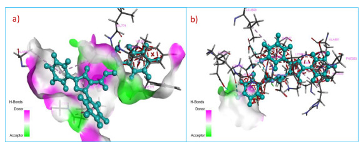
Figure2:Molecular dynamics simulation of (a) 3–(4–chlorophenyl)–5–(4–fluorophenyl)–4–phenyl–4, 5–dihydro–1,2,4–oxadiazole and (b) 3,5–bis–(4–chlorophenyl)–4–phenyl–4,5–dihydro–1,2,4–oxadiazole.
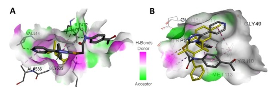
Figure3:Binding affinity performance for Nano synthesized (a) 3–(4–chlorophenyl)–5–(4–fluorophenyl)–4–phenyl–4, 5–dihydro–1, 2, 4–oxadiazole and (b) 3,5–bis–(4–chlorophenyl)–4–phenyl–4,5–dihydro–1,2,4–oxadiazole.
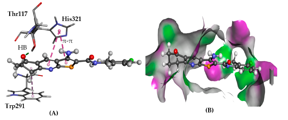
Figure 4: Pharmacokinetics (PK) of (a) 3–(4–chlorophenyl)–5–(4–fluorophenyl)–4–phenyl–4, 5–dihydro–1,2,4–oxadiazole and (b) 3,5–bis–(4–chlorophenyl)–4–phenyl–4,5–dihydro–1,2,4–oxadiazole.
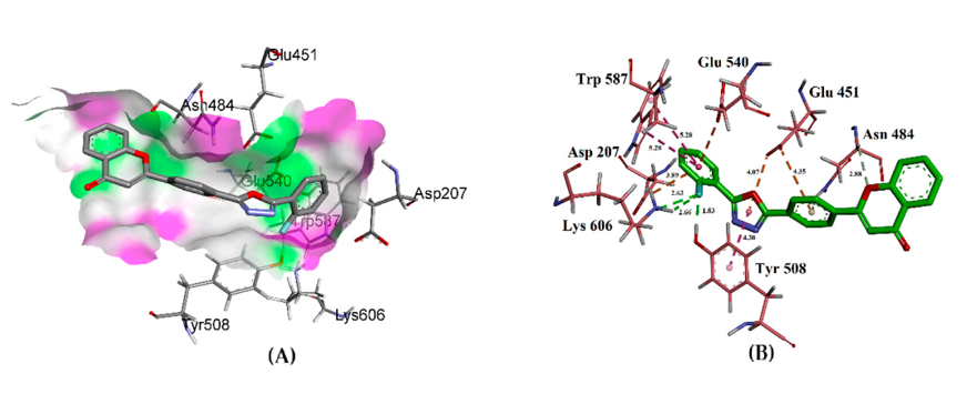
Figure5: Pharmacodynamics of (a) 3–(4–chlorophenyl)–5–(4–fluorophenyl)–4–phenyl–4, 5–dihydro–1,2,4–oxadiazole and (b) 3,5–bis–(4–chlorophenyl)–4–phenyl–4,5–dihydro–1,2,4–oxadiazole.
Figure 6:Quantitative Structure Activity Relationship (QSAR) and Quantitative Structure Properties Relationship (QSPR) of (a) 3–(4–chlorophenyl)–5–(4–fluorophenyl)–4–phenyl–4, 5–dihydro–1,2,4–oxadiazole and (b) 3,5–bis–(4–chlorophenyl)–4–phenyl–4,5–dihydro–1,2,4–oxadiazole.
Figure 7: Toxico and enzyme kinetics of (a) 3–(4–chlorophenyl)–5–(4–fluorophenyl)–4–phenyl–4, 5–dihydro–1, 2,4–oxadiazole and (b) 3,5–bis–(4–chlorophenyl)–4–phenyl–4,5–dihydro–1,2,4–oxadiazole.
Figure8: Effect of (a) 3–(4–chlorophenyl)–5–(4–fluorophenyl)–4–phenyl–4, 5–dihydro–1,2,4–oxadiazole and (b) 3,5–bis–(4–chlorophenyl)–4–phenyl–4,5–dihydro–1,2,4–oxadiazole on DNA/RNA in human cancer cells.
Figure9: Interaction of (a) 3–(4–chlorophenyl)–5–(4–fluorophenyl)–4–phenyl–4, 5–dihydro–1,2,4–oxadiazole and (b) 3,5–bis–(4–chlorophenyl)–4–phenyl–4,5–dihydro–1,2,4–oxadiazole with DNA/RNA in human cancer cells.
Results and Discussion
Absorption spectroscopy is one of the suitable methods for studying the structure of macromolecules. In order to investigate the spectroscopic properties of 3–(4–chlorophenyl)–5–(4–fluorophenyl)–4–phenyl–4, 5–dihydro–1, 2, 4–oxadiazole and 3, 5–bis–(4–chlorophenyl)–4–phenyl–4,5–dihydro–1,2,4–oxadiazole, first the absorption spectrum of this compound was drawn between 200 and 700 (nm) wavelength. The absorption spectrum of this anti–cancer Nano drug has a long absorption peak at 210 (nm) and several short absorption peaks at 208, 280, 320 and 439 (nm). Therefore, according to the absorption spectrum, the wavelength of 210 (nm) was chosen as the absorption index of 3–(4–chlorophenyl)–5–(4–fluorophenyl)–4–phenyl–4, 5–dihydro–1, 2, 4–oxadiazole and 3, 5–bis–(4–chlorophenyl)–4–phenyl–4, 5–dihydro–1, 2, 4–oxadiazole.In order to investigate the absorption changes of the samples incubated with 3–(4–chlorophenyl)–5–(4–fluorophenyl)–4–phenyl–4, 5–dihydro–1, 2, 4–oxadiazole and 3, 5–bis–(4–chlorophenyl)–4–phenyl–4, 5–dihydro–1, 2, 4–oxadiazole, UV/vis absorption spectrometry was used. Absorption spectroscopy is a useful method for studying the structure of different macromolecules, and on the other hand, this method is one of the simplest and most common methods for studying ligand–macromolecule interaction. First, the amount of absorption of standard samples of 3–(4–chlorophenyl)–5–(4–fluorophenyl)–4–phenyl–4,5–dihydro–1,2,4–oxadiazole and 3,5–bis–(4–chlorophenyl)–4–phenyl–4,5–dihydro–1,2,4–oxadiazole in each concentration is subtracted from the amount of absorption of samples treated with the same concentration of 3–(4–chlorophenyl)–5–(4–fluorophenyl)–4–phenyl–4,5–dihydro–1,2,4–oxadiazole and 3,5–bis–(4–chlorophenyl)–4–phenyl–4,5–dihydro–1,2,4–oxadiazole and the graph of changes in absorption of single–stranded DNA/RNA according to concentration. Obtained results show the changes in the absorbance of solutions resulting from the interaction of the anti–cancer Nano drug 3–(4–chlorophenyl)–5–(4–fluorophenyl)–4–phenyl–4,5–dihydro–1,2,4–oxadiazole and 3,5–bis–(4–chlorophenyl)–4–phenyl–4,5–dihydro–1,2,4–oxadiazole with single–stranded DNA/RNA at wavelengths of 260 (nm) (A)) and 210 (nm) (B) and as can be seen, the changes in single–stranded DNA/RNA absorption follow a similar process. In a comparative way, the changes in the absorbance of single–stranded DNA/RNA after incubation with 3–(4–chlorophenyl)–5–(4–fluorophenyl)–4–phenyl–4,5–dihydro–1,2,4–oxadiazole and 3,5–bis–(4–chlorophenyl)–4–phenyl–4,5–dihydro–1,2,4–oxadiazole at a wavelength of 260 (nm). In different concentrations, the amount of absorption of single–stranded DNA/RNA increases, and in higher concentrations, absorption also increases. While the decrease in absorption due to anti–cancer Nano drug interaction with DNA/RNA was not observed in any wavelength. As a result of the interaction of the anti–cancer Nano drug 3–(4–chlorophenyl)–5–(4–fluorophenyl)–4–phenyl–4,5–dihydro–1,2,4–oxadiazole and 3,5–bis–(4–chlorophenyl)–4–phenyl–4,5–dihydro–1,2,4–oxadiazole with single–stranded DNA/RNA at a wavelength of 210 (nm) in the presence of all concentrations of the anti–cancer Nano drug, absorption shows an increasing trend gives A closer observation shows that in low and high concentrations of 3–(4–chlorophenyl)–5–(4–fluorophenyl)–4–phenyl–4,5–dihydro–1,2,4–oxadiazole and 3,5–bis–(4–chlorophenyl)–4–phenyl–4,5–dihydro–1,2,4–oxadiazole, the absorption rate increases in different concentrations of 3–(4–chlorophenyl)–5–(4–fluorophenyl)–4–phenyl–4,5–dihydro–1,2,4–oxadiazole and 3,5–bis–(4–chlorophenyl)–4–phenyl–4,5–dihydro–1,2,4–oxadiazole.Next, in order to investigate the interaction of 3–(4–chlorophenyl)–5–(4–fluorophenyl)–4–phenyl–4,5–dihydro–1,2,4–oxadiazole and 3,5–bis–(4–chlorophenyl)–4–phenyl–4,5–dihydro–1,2,4–oxadiazole with single–stranded DNA/RNA, the fluorescence emission spectrum method was used. For this purpose, a certain concentration of single–stranded DNA/RNA was incubated separately with different concentrations of 3–(4–chlorophenyl)–5–(4–fluorophenyl)–4–phenyl–4,5–dihydro–1,2,4–oxadiazole and 3,5–bis–(4–chlorophenyl)–4–phenyl–4,5–dihydro–1,2,4–oxadiazole in the dark and at a temperature of 23 degrees Celsius for one hour and after the end of the time, the sample control and treated samples were excited at 258 (nm) wavelength and their absorption spectra were drawn. Then, the amount of fluorescence absorption of the standard samples of the anti–cancer Nano drug at each concentration was subtracted from the amount of absorption of the samples treated with the anti–cancer Nano drug at the same concentration, and the graph related to single–stranded DNA/RNA was drawn in the range of 500–550 (nm).At an excitation wavelength of 258 (nm) and in the absence of 3–(4–chlorophenyl)–5–(4–fluorophenyl)–4–phenyl–4,5–dihydro–1,2,4–oxadiazole and 3,5–bis–(4–chlorophenyl)–4–phenyl–4,5–dihydro–1,2,4–oxadiazole, single–stranded DNA/RNA has an absorption peak at 520 (nm), which corresponds to DNA/RNA chromophores (DNA/RNA bases) and by increasing the concentration of 3–(4–chlorophenyl)–5–(4–fluorophenyl)–4–phenyl–4,5–dihydro–1,2,4–oxadiazole and 3,5–bis–(4–chlorophenyl)–4–phenyl–4,5–dihydro–1,2,4–oxadiazole, the absorption rate of single–stranded DNA/RNA chromophores increases without any shift in the spectrum of single–stranded DNA/RNA. According to the fluorescence results, the I/Io×100 Io curve related to single–stranded DNA/RNA was also drawn against different anti–cancer Nano drug concentrations. Io is the fluorescence emission intensity of single–stranded DNA/RNA sample in the absence of 3–(4–chlorophenyl)–5–(4–fluorophenyl)–4–phenyl–4,5–dihydro–1,2,4–oxadiazole and 3,5–bis–(4–chlorophenyl)–4–phenyl–4,5–dihydro–1,2,4–oxadiazole and I is the fluorescence emission intensity of single–stranded DNA/RNA sample in the presence of different concentrations of 3–(4–chlorophenyl)–5–(4–fluorophenyl)–4–phenyl–4,5–dihydro–1,2,4–oxadiazole and 3,5–bis–(4–chlorophenyl)–4–phenyl–4,5–dihydro–1,2,4–oxadiazole. It can be seen that at a low concentration of 3–(4–chlorophenyl)–5–(4–fluorophenyl)–4–phenyl–4,5–dihydro–1,2,4–oxadiazole and 3,5–bis–(4–chlorophenyl)–4–phenyl–4,5–dihydro–1,2,4–oxadiazole (12.50 μg/ml), the affinity of the anti–cancer Nano drug for single–stranded DNA/RNA is almost low, and at higher concentrations (50–100 μg/ml) ml) of single–stranded DNA/RNA shows a greater affinity for 3–(4–chlorophenyl)–5–(4–fluorophenyl)–4–phenyl–4,5–dihydro–1,2,4–oxadiazole and 3,5–bis–(4–chlorophenyl)–4–phenyl–4,5–dihydro–1,2,4–oxadiazole. Stern–Volmer diagram shows the quenching effect of the anti–cancer Nano drug 3–(4–chlorophenyl)–5–(4–fluorophenyl)–4–phenyl–4, 5–dihydro–1,2,4–oxadiazole and 3,5–bis–(4–chlorophenyl)–4–phenyl–4,5–dihydro–1,2,4–oxadiazole. The slope of this graph is the Stern–Volmer constant, which is expressed in molar terms, and its value for single–stranded DNA/RNA is equal to 1.42×10³ M–1, which indicates that the anti–cancer Nano drug tends too much like single–stranded DNA/RNA. Obtained results show the binding constant of 3–(4–chlorophenyl)–5–(4–fluorophenyl)–4–phenyl–4,5–dihydro–1,2,4–oxadiazole and 3,5–bis–(4–chlorophenyl)–4–phenyl–4,5–dihydro–1,2,4–oxadiazole to single–stranded DNA/RNA M–1 = 68.35×10³Ka and the number of binding site n = 72.In order to study the effect of 3–(4–chlorophenyl)–5–(4–fluorophenyl)–4–phenyl–4,5–dihydro–1,2,4–oxadiazole and 3,5–bis–(4–chlorophenyl)–4–phenyl–4,5–dihydro–1,2,4–oxadiazole on the second structure of single–stranded DNA/RNA molecule, the circular dipole (CD) method was used in the near ultraviolet range. For this purpose, single–stranded DNA/RNA was incubated with different concentrations of 3–(4–chlorophenyl)–5–(4–fluorophenyl)–4–phenyl–4,5–dihydro–1,2,4–oxadiazole and 3,5–bis–(4–chlorophenyl)–4–phenyl–4,5–dihydro–1,2,4–oxadiazole at room temperature and away from light for one hour. After a certain time, the CD spectrum of the samples was drawn in the absence of single–stranded DNA/RNA. Then the CD spectrum of different anti–cancer Nano drug concentrations in each concentration was subtracted from the spectrum of samples treated with 3–(4–chlorophenyl)–5–(4–fluorophenyl)–4–phenyl–4,5–dihydro–1,2,4–oxadiazole and 3,5–bis–(4–chlorophenyl)–4–phenyl–4,5–dihydro–1,2,4–oxadiazole at the same concentration. As seen in the Figures (1–9), the spectrum of single–stranded DNA/RNA in the absence of 3–(4–chlorophenyl)–5–(4–fluorophenyl)–4–phenyl–4,5–dihydro–1,2,4–oxadiazole and 3,5–bis–(4–chlorophenyl)–4–phenyl–4,5–dihydro–1,2,4–oxadiazole has two positive regions, one at 220 (nm) and the other at 275 (nm), and a negative region at 245 (nm). Increasing the concentration of 3–(4–chlorophenyl)–5–(4–fluorophenyl)–4–phenyl–4, 5–dihydro–1,2,4–oxadiazole and 3,5–bis–(4–chlorophenyl)–4–phenyl–4,5–dihydro–1,2,4–oxadiazole affects the ellipticity of the single–stranded DNA/RNA molecule in all regions including at 275, 245 and 220 (nm). At 275 (nm) and in the presence of the anti–cancer Nano drug, the ellipticity increases and shifts to shorter wavelengths. At 245 (nm), with the addition of 3–(4–chlorophenyl)–5–(4–fluorophenyl)–4–phenyl–4,5–dihydro–1,2,4–oxadiazole and 3,5–bis–(4–chlorophenyl)–4–phenyl–4,5–dihydro–1,2,4–oxadiazole, the ellipticity becomes more positive and the graph remains unchanged in the wavelength, and finally, at 220 (nm), in addition to increasing the ellipticity, there is some shift towards shorter wavelengths.
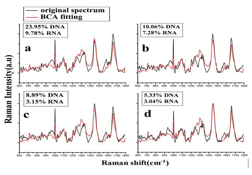
The anti–tumor activity of natural and synthetic oxadiazole derivatives has been widely investigated and it has been proven that oxadiazoles, depending on their structure, can act on different tumor cells by different mechanisms of action such as telomerase enzyme inhibition, protein activity inhibition Kinase and downregulation of oncogene expression also induce caspase mediated apoptosis, and suppress cancer cell proliferation by arresting the cell cycle in the G0/G1 phase, G2/M phase. DNA/RNA in the nucleus of eukaryotic cells is naked during replication and may be targeted by many different compounds or enzymes, but it is not naked in the usual state and is connected to histone and non–histone proteins, which together make the nucleoprotein composition Creates the name chromatin. On the other hand, in the structure of chromatin, DNA/RNA forms complex histone octamer and nucleosome. The DNA/RNA compounds in the cell nucleus are among the most important targets of many anti–cancer Nano drugs after they enter the cell nucleus, and so far, many studies have been conducted on the effect of anti–tumor Nano drugs on the structure of chromatin and DNA/RNA. There is no information about the effect of 3–(4–chlorophenyl)–5–(4–fluorophenyl)–4–phenyl–4,5–dihydro–1,2,4–oxadiazole and 3,5–bis–(4–chlorophenyl)–4–phenyl–4,5–dihydro–1,2,4–oxadiazole on single–stranded DNA/RNA, and there is no available data about the mentioned compound on DNA/RNA structure. For this reason, it was decided that the effect of this anti–cancer Nano drug on soluble single–stranded DNA/RNA was investigated and the results of its interaction with single–stranded DNA/RNA were investigated. For this purpose, a constant concentration of single–stranded DNA/RNA with different concentrations of 3–(4–chlorophenyl)–5–(4–fluorophenyl)–4–phenyl–4,5–dihydro–1,2,4–oxadiazole and 3,5–bis–(4–chlorophenyl)–4–phenyl–4,5–dihydro–1,2,4–oxadiazole was incubated at room temperature and away from light, and after the desired period of time, various spectroscopic studies were performed on them. The results obtained from the increase in absorption of single–stranded DNA/RNA at 210 and 260 (nm) depend on the concentration and indicate the participation of phosphate groups and DNA/RNA bases in the interaction with the anti–cancer Nano drug. At high concentrations of 3–(4–chlorophenyl)–5–(4–fluorophenyl)–4–phenyl–4,5–dihydro–1,2,4–oxadiazole and 3,5–bis–(4–chlorophenyl)–4–phenyl–4,5–dihydro–1,2,4–oxadiazole, the structure of single–stranded DNA/RNA is opened and as a result, the absorption rate increases. The increased intensity of absorption in the case of single–stranded DNA/RNA indicates the greater tendency of the anti–cancer Nano drug to bind to single–stranded DNA/RNA. Fluorescence spectroscopy is more efficient, sensitive and more complex than absorption spectroscopy. Using the fluorescence spectroscopy method, important information about the macromolecule structure can be obtained. The increase in the fluorescence intensity of single–stranded DNA/RNA indicates the opening of DNA/RNA chromophores in the presence of 3–(4–chlorophenyl)–5–(4–fluorophenyl)–4–phenyl–4,5–dihydro–1,2,4–oxadiazole and 3,5–bis–(4–chlorophenyl)–4–phenyl–4,5–dihydro–1,2,4–oxadiazole, and the linearity of the Stern–Volmer curve also confirms this. The I?–I/I?×100 curve of single–stranded DNA/RNA also shows the high affinity of 3–(4–chlorophenyl)–5–(4–fluorophenyl)–4–phenyl–4, 5–dihydro–1, 2, 4–oxadiazole and 3,5–bis–(4–chlorophenyl)–4–phenyl–4,5–dihydro–1,2,4–oxadiazole to single–stranded DNA/RNA. On the other hand, the opening of single–stranded DNA/RNA chromophores can be attributed to the intercalation of 3–(4–chlorophenyl)–5–(4–fluorophenyl)–4–phenyl–4, 5–dihydro–1, 2, 4–oxadiazole and 3,5–bis–(4–chlorophenyl)–4–phenyl–4,5–dihydro–1,2,4–oxadiazole inside these structures. In a study where oxadiazole derivatives such as 3–(4–chlorophenyl)–5–(4–fluorophenyl)–4–phenyl–4,5–dihydro–1,2,4–oxadiazole and 3,5–bis–(4–chlorophenyl)–4–phenyl–4,5–dihydro–1,2,4–oxadiazole 35 (Ksv=25.61×10³ M–1) were performed on bovine serum albumin (BSA) and also in another study that investigated another oxadiazole derivative called 4–methyl–7–hydroxy oxadiazole was studied in the presence of BSA and HSA, Ksv is equal to 1.80×10³ M–1 and 4.97×10³ M–1, respectively, which means that 3–(4–chlorophenyl)–5–(4–fluorophenyl)–4–phenyl–4,5–dihydro–1,2,4–oxadiazole and 3,5–bis–(4–chlorophenyl)–4–phenyl–4,5–dihydro–1,2,4–oxadiazole (M–1 = 103×42/1Ksv) has a lower Stern–Volmer equation constant than these oxadiazole derivatives. In another study where the effect of oxadiazole was performed in the presence of ethidium bromide and acridine orange, the Ksv was 0.21×10³ M–1 and 0.56×10³ M–1, respectively, which is the Ksv corresponding to 3–(4–chlorophenyl)–5–(4–fluorophenyl)–4–phenyl–4,5–dihydro–1,2,4–oxadiazole and 3,5–bis–(4–chlorophenyl)–4–phenyl–4,5–dihydro–1,2,4–oxadiazole are less. CD spectroscopy is a powerful method in studying the conformational properties of single–stranded DNA/RNA molecules and is used in studying the interaction of nucleic acids with proteins and ligands. The change in the ellipticity of the DNA/RNA molecule in the region of 245 (nm) indicates the change in the orientation of B–DNA/RNA. In addition, anti–cancer Nano drug interaction at 220 and 275 (nm) causes an increase in ellipticity. In general, the changes at 220, 245 and 275 (nm) indicate the effect of the anti–cancer Nano drug on the stacking of bases, which causes a change in the B conformation in the DNA/RNA structure and a decrease in the second structures of the DNA/RNA molecule, and possibly changes B–DNA/RNA to A or C–DNA/RNA(Figure 10).
Figure 10: Raman spectra of DNA/RNA linked to 3–(4–chlorophenyl)–5–(4–fluorophenyl)–4–phenyl–4,5–dihydro–1,2,4–oxadiazole and 3,5–bis–(4–chlorophenyl)–4–phenyl–4,5–dihydro–1,2,4–oxadiazole in (a and b) human normal cells and (c and d) human cancer cells.
Conclusion
According to the results of the interaction of 3–(4–chlorophenyl)–5–(4–fluorophenyl)–4–phenyl–4,5–dihydro–1,2,4–oxadiazole and 3,5–bis–(4–chlorophenyl)–4–phenyl–4,5–dihydro–1,2,4–oxadiazole with single–stranded DNA/RNA, which indicates the high tendency of the anti–cancer Nano drug to bind to single–stranded DNA/RNA, the involvement of single–stranded DNA/RNA is probably one of the most important The most important targets in chromatin structure are 3–(4–chlorophenyl)–5–(4–fluorophenyl)–4–phenyl–4,5–dihydro–1,2,4–oxadiazole and 3,5–bis–(4–chlorophenyl)–4–phenyl–4,5–dihydro–1,2,4–oxadiazole.
Acknowledgement
This study was supported by the Cancer Research Institute (CRI) Project of Scientific Instrument and Equipment Development, the National Natural Science Foundation of the United Sates, the International Joint BioSpectroscopy Core Research Laboratory (BCRL) Program supported by the California South University (CSU), and the Key project supported by the American International Standards Institute (AISI), Irvine, California, USA.
References
- J Med Oncol. 1: 1.
- “A Modern Ethnomedicinal Technique for Transformation, Prevention and Treatment of Human Malignant Gliomas Tumors into Human Benign Gliomas Tumors under Synchrotron Radiation”. Am J Ethnomed 1:10.
- J Med Chem Toxicol 2: 1–5.
- “Investigation of Medical, Medicinal, Clinical and Pharmaceutical Applications of Estradiol, Mestranol (Norlutin), Norethindrone (NET), Norethisterone Acetate (NETA), Norethisterone Enanthate (NETE) and Testosterone Nanoparticles as Biological Imaging, Cell Labeling, Anti–Microbial Agents and Anti–Cancer Nano Drugs in Nanomedicines Based Drug Delivery Systems for Anti–Cancer Targeting and Treatment”, Parana Journal of Science and Education 3: 10–19.
- “A Comparative Computational and Experimental Study on Different Vibrational Biospectroscopy Methods, Techniques and Applications for Human Cancer Cells in Tumor Tissues Simulation, Modeling, Research, Diagnosis and Treatment”. Open J Anal Bioanal Chem 1: 014–020.
- “Combination of DNA/RNA Ligands and Linear/Non–Linear Visible–Synchrotron Radiation–Driven N–Doped Ordered Mesoporous Cadmium Oxide (CdO) Nanoparticles Photocatalysts Channels Resulted in an Interesting Synergistic Effect Enhancing Catalytic Anti–Cancer Activity”. Enz Eng 6: 1.
- “Modern Approaches in Designing Ferritin, Ferritin Light Chain, Transferrin, Beta–2 Transferrin and Bacterioferritin–Based Anti–Cancer Nano Drugs Encapsulating Nanosphere as DNA–Binding Proteins from Starved Cells (DPS)”. Mod Appro Drug Des 1: 000504.
- “Potency of Human Interferon β–1a and Human Interferon β–1b in Enzymotherapy, Immunotherapy, Chemotherapy, Radiotherapy, Hormone Therapy and Targeted Therapy of Encephalomyelitis Disseminate/Multiple Sclerosis (MS) and Hepatitis A, B, C, D, E, F and G Virus Enter and Targets Liver Cells”. J Proteomics Enzymol 6: 1.
- “Transport Therapeutic Active Targeting of Human Brain Tumors Enable Anti–Cancer Nanodrugs Delivery across the Blood–Brain Barrier (BBB) to Treat Brain Diseases Using Nanoparticles and Nanocarriers under Synchrotron Radiation”. J Pharm Pharmaceutics 4: 1–5.
- C. Brown, “Combinatorial Therapeutic Approaches to DNA/RNA and Benzylpenicillin (Penicillin G), Fluoxetine Hydrochloride (Prozac and Sarafem), Propofol (Diprivan), Acetylsalicylic Acid (ASA) (Aspirin), Naproxen Sodium (Aleve and Naprosyn) and Dextromethamphetamine Nanocapsules with Surface Conjugated DNA/RNA to Targeted Nano Drugs for Enhanced Anti–Cancer Efficacy and Targeted Cancer Therapy Using Nano Drugs Delivery Systems”. Ann Adv Chem 1: 061–069.
- Heidari, (2017) “High–Resolution Simulations of Human Brain Cancer Translational Nano Drugs Delivery Treatment Process under Synchrotron Radiation”. J Transl Res 1: 1–3.
- “Investigation of Anti–Cancer Nano Drugs’ Effects’ Trend on Human Pancreas Cancer Cells and Tissues Prevention, Diagnosis and Treatment Process under Synchrotron and X–Ray Radiations with the Passage of Time Using Mathematica”. Current Trends Anal Bioanal Chem 1: 36–41.
- “Pros and Cons Controversy on Molecular Imaging and Dynamics of Double–Standard DNA/RNA of Human Preserving Stem Cells–Binding Nano Molecules with Androgens/Anabolic Steroids (AAS) or Testosterone Derivatives through Tracking of Helium–4 Nucleus (Alpha Particle) Using Synchrotron Radiation”. Arch Biotechnol Biomed 1: 67–100.
- “Visualizing Metabolic Changes in Probing Human Cancer Cells and Tissues Metabolism Using Vivo 1H or Proton NMR, 13C NMR, 15N NMR and 31P NMR Spectroscopy and Self–Organizing Maps under Synchrotron Radiation”. SOJ Mater Sci Eng 5: 1–6.
- “Cavity Ring–Down Spectroscopy (CRDS), Circular Dichroism Spectroscopy, Cold Vapour Atomic Fluorescence Spectroscopy and Correlation Spectroscopy Comparative Study on Malignant and Benign Human Cancer Cells and Tissues with the Passage of Time under Synchrotron Radiation”. Enliven: Challenges Cancer Detect Ther 4: 001.
- “Laser Spectroscopy, Laser–Induced Breakdown Spectroscopy and Laser–Induced Plasma Spectroscopy Comparative Study on Malignant and Benign Human Cancer Cells and Tissues with the Passage of Time under Synchrotron Radiation”. Int J Hepatol Gastroenterol 3: 79–84.
- “Time–Resolved Spectroscopy and Time–Stretch Spectroscopy Comparative Study on Malignant and Benign Human Cancer Cells and Tissues with the Passage of Time under Synchrotron Radiation”. Enliven: Pharmacovigilance and Drug Safety 4: 001.
- “Overview of the Role of Vitamins in Reducing Negative Effect of Decapeptyl (Triptorelin Acetate or Pamoate Salts) on Prostate Cancer Cells and Tissues in Prostate Cancer Treatment Process through Transformation of Malignant Prostate Tumors into Benign Prostate Tumors under Synchrotron Radiation”. Open J Anal Bioanal Chem 1: 21–26.
- “Electron Phenomenological Spectroscopy, Electron Paramagnetic Resonance (EPR) Spectroscopy and Electron Spin Resonance (ESR) Spectroscopy Comparative Study on Malignant and Benign Human Cancer Cells and Tissues with the Passage of Time under Synchrotron Radiation”. Austin J Anal Pharm Chem 4: 1091.
- “Therapeutic Nanomedicine Different High–Resolution Experimental Images and Computational Simulations for Human Brain Cancer Cells and Tissues Using Nanocarriers Deliver DNA/RNA to Brain Tumors under Synchrotron Radiation with the Passage of Time Using Mathematica and MATLAB”. Madridge J Nano Tech. Sci 2: 77–83.
- “A Consensus and Prospective Study on Restoring Cadmium Oxide (CdO) Nanoparticles Sensitivity in Recurrent Ovarian Cancer by Extending the Cadmium Oxide (CdO) Nanoparticles–Free Interval Using Synchrotron Radiation Therapy as Antibody–Drug Conjugate for the Treatment of Limited–Stage Small Cell Diverse Epithelial Cancers” Cancer Clin Res Rep 1: 001.
- “A Novel and Modern Experimental Imaging and Spectroscopy Comparative Study on Malignant and Benign Human Cancer Cells and Tissues with the Passage of Time under White Synchrotron Radiation”. Cancer Sci Res Open Access 4: 1–8.
- “Different High–Resolution Simulations of Medical, Medicinal, Clinical, Pharmaceutical and Therapeutics Oncology of Human Breast Cancer Translational Nano Drugs Delivery Treatment Process under Synchrotron and X–Ray Radiations”. J Oral Cancer Res 1: 12–17.
- “Vibrational Decihertz (dHz), Centihertz (cHz), Millihertz (mHz), Microhertz (μHz), Nanohertz (nHz), Picohertz (pHz), Femtohertz (fHz), Attohertz (aHz), Zeptohertz (zHz) and Yoctohertz (yHz) Imaging and Spectroscopy Comparative Study on Malignant and Benign Human Cancer Cells and Tissues under Synchrotron Radiation”. International Journal of Biomedicine 7: 335–340.
- “Force Spectroscopy and Fluorescence Spectroscopy Comparative Study on Malignant and Benign Human Cancer Cells and Tissues with the Passage of Time under Synchrotron Radiation”.EC Cancer 2: 239–246.
- “Photoacoustic Spectroscopy, Photoemission Spectroscopy and Photothermal Spectroscopy Comparative Study on Malignant and Benign Human Cancer Cells and Tissues with the Passage of Time under Synchrotron Radiation”. BAOJ Cancer Res Ther 3: 45–052.
- “J–Spectroscopy, Exchange Spectroscopy (EXSY), Nuclear Overhauser Effect Spectroscopy (NOESY) and Total Correlation Spectroscopy (TOCSY) Comparative Study on Malignant and Benign Human Cancer Cells and Tissues under Synchrotron Radiation”. EMS Eng Sci J 1: 006–013.
- “Neutron Spin Echo Spectroscopy and Spin Noise Spectroscopy Comparative Study on Malignant and Benign Human Cancer Cells and Tissues with the Passage of Time under Synchrotron Radiation”. Int J Biopharm Sci 1: 103–107.
- “Vibrational Decahertz (daHz), Hectohertz (hHz), Kilohertz (kHz), Megahertz (MHz), Gigahertz (GHz), Terahertz (THz), Petahertz (PHz), Exahertz (EHz), Zettahertz (ZHz) and Yottahertz (YHz) Imaging and Spectroscopy Comparative Study on Malignant and Benign Human Cancer Cells and Tissues under Synchrotron Radiation”. Madridge J Anal Sci Instrum 2: 41–46.
- “Two–Dimensional Infrared Correlation Spectroscopy, Linear Two–Dimensional Infrared Spectroscopy and Non–Linear Two–Dimensional Infrared Spectroscopy Comparative Study on Malignant and Benign Human Cancer Cells and Tissues under Synchrotron Radiation with the Passage of Time” J Mater Sci Nanotechnol 6: 101.
- “Fourier Transform Infrared (FTIR) Spectroscopy, Near–Infrared Spectroscopy (NIRS) and Mid–Infrared Spectroscopy (MIRS) Comparative Study on Malignant and Benign Human Cancer Cells and Tissues under Synchrotron Radiation with the Passage of Time”. Int J Nanotechnol Nanomed 3: 1–6.
- ) “Infrared Photo Dissociation Spectroscopy and Infrared Correlation Table Spectroscopy Comparative Study on Malignant and Benign Human Cancer Cells and Tissues under Synchrotron Radiation with the Passage of Time”. Austin Pharmacol Pharm 3: 1011.
- “Novel and Transcendental Prevention, Diagnosis and Treatment Strategies for Investigation of Interaction among Human Blood Cancer Cells, Tissues, Tumors and Metastases with Synchrotron Radiation under Anti–Cancer Nano Drugs Delivery Efficacy Using MATLAB Modeling and Simulation”. Madridge J Nov Drug Res 1: 18–24.
- “Comparative Study on Malignant and Benign Human Cancer Cells and Tissues with the Passage of Time under Synchrotron Radiation”. Open Access J Trans Med Res 2: 26-32.
- ) “Planting of Jaboticaba Trees for Landscape Repair of Degraded Area”. Landscape Architecture and Regional Planning 3: 1–9.
- “Fluorescence Spectroscopy, Phosphorescence Spectroscopy and Luminescence Spectroscopy Comparative Study on Malignant and Benign Human Cancer Cells and Tissues under Synchrotron Radiation with the Passage of Time”. SM J Clin. Med. Imaging 4: 1018.
- “Nuclear Inelastic Scattering Spectroscopy (NISS) and Nuclear Inelastic Absorption Spectroscopy (NIAS) Comparative Study on Malignant and Benign Human Cancer Cells and Tissues under Synchrotron Radiation. Int J Pharm Sci 2: 1–14.
- “X–Ray Diffraction (XRD), Powder X–Ray Diffraction (PXRD) and Energy–Dispersive X–Ray Diffraction (EDXRD) Comparative Study on Malignant and Benign Human Cancer Cells and Tissues under Synchrotron Radiation”. J Oncol Res 2: 1–14.
- “Correlation Two–Dimensional Nuclear Magnetic Resonance (NMR) (2D–NMR) (COSY) Imaging and Spectroscopy Comparative Study on Malignant and Benign Human Cancer Cells and Tissues under Synchrotron Radiation”. EMS Can Sci 1:1–001.
- “Thermal Spectroscopy, Photothermal Spectroscopy, Thermal Microspectroscopy, Photothermal Microspectroscopy, Thermal Macrospectroscopy and Photothermal Macrospectroscopy Comparative Study on Malignant and Benign Human Cancer Cells and Tissues with the Passage of Time under Synchrotron Radiation”. SM J Biometrics Biostat 3: 1024.
- “A Modern and Comprehensive Experimental Biospectroscopic Comparative Study on Human Common Cancers’ Cells, Tissues and Tumors before and after Synchrotron Radiation Therapy”. Open Acc J Oncol Med 1.
- “Heteronuclear Correlation Experiments Such as Heteronuclear Single–Quantum Correlation Spectroscopy (HSQC), Heteronuclear Multiple–Quantum Correlation Spectroscopy (HMQC) and Heteronuclear Multiple–Bond Correlation Spectroscopy (HMBC) Comparative Study on Malignant and Benign Human Endocrinology and Thyroid Cancer Cells and Tissues under Synchrotron Radiation” J Endocrinol Thyroid Res 3: 555-603.
- “Nuclear Resonance Vibrational Spectroscopy (NRVS), Nuclear Inelastic Scattering Spectroscopy (NISS), Nuclear Inelastic Absorption Spectroscopy (NIAS) and Nuclear Resonant Inelastic X–Ray Scattering Spectroscopy (NRIXSS) Comparative Study on Malignant and Benign Human Cancer Cells and Tissues under Synchrotron Radiation”. Int J Bioorg Chem Mol Biol 6: 1–5.
- “A Novel and Modern Experimental Approach to Vibrational Circular Dichroism Spectroscopy and Video Spectroscopy Comparative Study on Malignant and Benign Human Cancer Cells and Tissues with the Passage of Time under White and Monochromatic Synchrotron Radiation”. Glob J Endocrinol Metab 1: 514–519.
- “Pros and Cons Controversy on Heteronuclear Correlation Experiments Such as Heteronuclear Single–Quantum Correlation Spectroscopy (HSQC), Heteronuclear Multiple–Quantum Correlation Spectroscopy (HMQC) and Heteronuclear Multiple–Bond Correlation Spectroscopy (HMBC) Comparative Study on Malignant and Benign Human Cancer Cells and Tissues under Synchrotron Radiation”. EMS Pharma J 1: 2-8.
- “A Modern Comparative and Comprehensive Experimental Biospectroscopic Study on Different Types of Infrared Spectroscopy of Malignant and Benign Human Cancer Cells and Tissues with the Passage of Time under Synchrotron Radiation”. J Analyt Molecul Tech 3: 8.
- “Investigation of Cancer Types Using Synchrotron Technology for Proton Beam Therapy: An Experimental Biospectroscopic Comparative Study”. European Modern Studies Journal 2: 13-29.
- “Saturated Spectroscopy and Unsaturated Spectroscopy Comparative Study on Malignant and Benign Human Cancer Cells and Tissues with the Passage of Time under Synchrotron Radiation”. Imaging J Clin Medical Sci 5: 1-7.
- Small–Angle Neutron Scattering (SANS) and Wide–Angle X–Ray Diffraction (WAXD) Comparative Study on Malignant and Benign Human Cancer Cells and Tissues under Synchrotron Radiation”. Int J Bioorg Chem Mol Biol 6: 1-6.
- “Investigation of Bladder Cancer, Breast Cancer, Colorectal Cancer, Endometrial Cancer, Kidney Cancer, Leukemia, Liver, Lung Cancer, Melanoma, Non–Hodgkin Lymphoma, Pancreatic Cancer, Prostate Cancer, Thyroid Cancer and Non–Melanoma Skin Cancer Using Synchrotron Technology for Proton Beam Therapy: An Experimental Biospectroscopic Comparative Study”. Ther Res Skin Dis 1.
- “Attenuated Total Reflectance Fourier Transform Infrared (ATR–FTIR) Spectroscopy, Micro–Attenuated Total Reflectance Fourier Transform Infrared (Micro–ATR–FTIR) Spectroscopy and Macro–Attenuated Total Reflectance Fourier Transform Infrared (Macro–ATR–FTIR) Spectroscopy Comparative Study on Malignant and Benign Human Cancer Cells and Tissues under Synchrotron Radiation with the Passage of Time”. International Journal of Chemistry Papers 2: 1–12.
- “Mössbauer Spectroscopy, Mössbauer Emission Spectroscopy and 57Fe Mössbauer Spectroscopy Comparative Study on Malignant and Benign Human Cancer Cells and Tissues under Synchrotron Radiation”. Acta Scientific Cancer Biology 2: 17–20.
- “Comparative Study on Malignant and Benign Human Cancer Cells and Tissues under Synchrotron Radiation with the Passage of Time”. Organic & Medicinal Chem IJ 6: 555676.
- “Correlation Spectroscopy, Exclusive Correlation Spectroscopy and Total Correlation Spectroscopy Comparative Study on Malignant and Benign Human AIDS–Related Cancers Cells and Tissues with the Passage of Time under Synchrotron Radiation”. Int J Bioanal Biomed 2: 1-7.
- “Biomedical Instrumentation and Applications of Biospectroscopic Methods and Techniques in Malignant and Benign Human Cancer Cells and Tissues Studies under Synchrotron Radiation and Anti–Cancer Nano Drugs Delivery”. Am J Nanotechnol Nanomed 1: 1-09.
- “Vivo 1H or Proton NMR, 13C NMR, 15N NMR and 31P NMR Spectroscopy Comparative Study on Malignant and Benign Human Cancer Cells and Tissues under Synchrotron Radiation”. Ann Biomet Biostat 1: 1001.
- Ann Cardiovasc Surg 1: 1-006.
- “Adsorption Isotherms and Kinetics of Multi–Walled Carbon Nanotubes (MWCNTs), Boron Nitride Nanotubes (BNNTs), Amorphous Boron Nitride Nanotubes (a–BNNTs) and Hexagonal Boron Nitride Nanotubes (h–BNNTs) for Eliminating Carcinoma, Sarcoma, Lymphoma, Leukemia, Germ Cell Tumor and Blastoma Cancer Cells and Tissues”. Clin Med Rev Case Rep 5: 201.
- ”. Acta Scientific Pharmaceutical Sciences 5: 30–35.
- A Heidari (2018) “Small–Angle X–Ray Scattering (SAXS), Ultra–Small Angle X–Ray Scattering (USAXS), Fluctuation X–Ray Scattering (FXS), Wide–Angle X–Ray Scattering (WAXS), Grazing–Incidence Small–Angle X–Ray Scattering (GISAXS), Grazing–Incidence Wide–Angle X–Ray Scattering (GIWAXS), Small–Angle Neutron Scattering (SANS), Grazing–Incidence Small–Angle Neutron Scattering (GISANS), X–Ray Diffraction (XRD), Powder X–Ray Diffraction (PXRD), Wide–Angle X–Ray Diffraction (WAXD), Grazing–Incidence X–Ray Diffraction (GIXD) and Energy–Dispersive X–Ray Diffraction (EDXRD) Comparative Study on Malignant and Benign Human Cancer Cells and Tissues under Synchrotron Radiation”. Oncol Res Rev 1: 1–10.
- A Heidari (2018) “A Modern and Comprehensive Investigation of Inelastic Electron Tunneling Spectroscopy (IETS) and Scanning Tunneling Spectroscopy on Malignant and Benign Human Cancer Cells, Tissues and Tumors through Optimizing Synchrotron Microbeam Radiotherapy for Human Cancer Treatments and Diagnostics: An Experimental Biospectroscopic Comparative.