Intra Oral Verrucous Carcinoma with Foci of Invasive Squamous Cell Carcinoma – A Rare Case Report from Egypt.
Mohamed Said Hamed1, Amr El Swify1, Magda Hassan2, Anas Mahmoud Taalab3*
1 Professor of Oral and Maxillofacial Surgery , Faculty of Dentistry , Suez Canal University.
2 Professorof Oral Pathology , Faculty of Dentistry , Suez Canal University.
3 Teaching Assistant at Oral and Maxillofacial Surgery Department, Faculty of Dentistry, Suez Canal University.
*Corresponding Author: Anas Mahmoud Taalab, Teaching Assistant at Oral and Maxillofacial Surgery Department, Faculty of Dentistry, Suez Canal University, Egypt, TEL: +20 64 3200395; FAX: +20 64 3200395; E-mail: anas.ta3lab@gmail.com
Citation: Mohamed Said Hamed, Amr El Swify, Magda Hassan, Anas Mahmoud Taalab (2019) Intra Oral Verrucous Carcinoma with Foci of Invasive Squamous Cell Carcinoma – A Rare Case Report from Egypt. Medcina Intern 2019 3: 140
Copyright: © 2019 Anas Mahmoud Taalab, et al. This is an open-access article distributed under the terms of the Creative Commons Attribution License, which permits unrestricted use, distribution, and reproduction in any medium, provided the original author and source are credited
Received date: December 26, 2019; Accepted date: December 29, 2019; Published date: December 31, 2019.
Abstract
Verrucous carcinoma (VC) is an uncommon type of low-grade, well differentiated andnon-invasive squamous cell carcinoma (SCC) with specific clinical and histological features. We report a case of a 65 years old female presenting a papillary, easily bleeding painful, reddish with leukoplastic lesion in maxillary alveolar ridge measuring approximately 5cm diameter, with 2 months evolution.
Histological examination of the incisional biopsy revealed proliferation of well-differentiated squamous epithelium forming high exophytic hyperparakeratinized papillary projections with variable width of rete ridges .par keratin plugs extended from the surface to about 2/3 of epithelial thickness.
Mixed inflammatory cellular infiltrate extended to the non-keratinized part of the epithelium .most of the hyperplastic epithelium is normal appearing, except for increased tendency for keratinization of the prickle cells
In focal areas, the rete ridges were more narrower and showed atypical features as loss of polarity of basal cells, hyperplastic basal cell layer and hyperchoromatism.
Extending from these ridges multiple epithelium nests and islands invading the connective tissue, showing more dysplastic criteria as cellular and nuclear pleomorphism, Para keratin pearl formation increased tendency for individual cell keratinization ,swirling pattern ,prominent nucleoli and minimal mitotic figures.
Diagnosis: is well to moderately differentiated SCC on top of VC. The patient underwent surgery with a safety margin and is under follow-up.
Keywords:
Carcinoma, Verrucous. Carcinoma, Squamous Cell
Introduction
Verrucous carcinoma (VC) is a raretumor. It's is a well-recognized variant of the squamous cell carcinoma (SCC) of the oral cavity with unique morphology, characterized by exophytic appearance, presenting locally destructive growth but no metastasis tendency. It was first identified as a clinical and histologic entity by Ackermann in 1948.
Various names are used in the literature to describe this entity, including Ackerman’s tumor, Buschke-Loewenstein tumor, florid oral papillomatosis, epitheliomacuniculatum, and carcinoma cuniculatum.
It appears as a painless, thick white plaque resembling a cauliflower. The most common sites of oral mucosal involvement include the buccal mucosa, followed by the mandibular alveolar crest, gingiva, and tongue.
The histological presentation of VC is well defined, and it is characterized by epithelial exophytic projections forming high sharp ridges with keratin-filled invaginations, and downgrowth as blunt papillas that seem to compress the underlying connective tissue, with minimal or no cytological atypia [1,2].
The non-invasive growth pattern and minimal or no cytological atipia of tumor cells are topical features to separate these tumors from classical invasive oral squamous cell carcinoma [2].
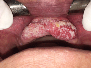
However, “hybrid” lesions comprised of typical VC associated with SCC have been previously reported [3]. The hybrid VC associated with SCC can represent up to 20% of these tumors in the oral cavity, and the differential diagnosis is considered an important diagnostic dillema, since some studies report that these lesions behaves like SCC rather than VC regarding their metastatic tendency [4], whereas others suggest that the clinical behavior of these tumors more closely matches pure VC [3]. Figure 1.

Figure 1: Clinical presentation of the lesion as an extensive painfull verrucous leukoplastic lesion, with 50 mm in the largest diameter, located in the anterior part of maxillary ridge.
As there are only some few reports of VC in the literature, we describe a case of this hybrid lesion in a 65 years old female patient.
Case Report
A 65-year-old white female was referred to a faculty of dentistry SCU with a chief complaint of a painful white papillary mass at the area of premaxilla . Intraoral examinations revealed an extensive verrucous leukoplastic lesion, with 5.0 cm in the largest diameter, symptomatic, located in the anterior part of maxillary ridge (Fig. 1), with 2 months evolution.
The presumptive diagnoses were verrucous carcinoma × benign verrucous hyperplasia × squamous cell carcinoma. Incisional biopsy was performed and the tissue sample was sent to histological examinations.
Histopathologic finding
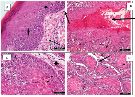
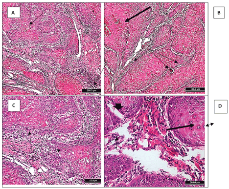
On histologic review of the submitted tissue, the overall presentation was hyperparakeratinised, hyperplastic stratified squamous epithelium with papillary projections that were filled with massive amount of parakeratin extensions. The safety margin of the lesion was parakeratinized stratified epithelium, lacking the normal gingival rete ridges’ appearance, which were broader, with signs of dysplasia as loss of basal cell polarity, basal cell hyperplasia, cellular and nuclear pleomorphism (Figure 2A). The papillary projections of the main lesion had elongated downward extensions, most of them had irregular budding rete processes (Figure 2B). Higher power of these ends revealed signs of dysplasia as reversed nuclear/ cytoplasmic ratio, basal cell hyperplasia and significant tendency for keratinization of the supra-basal cells (Figure 2C). At focal areas there were invasion of dysplastic epithelium towards inflamed connective tissue, forming nests and sheets, where most suprabasal cells had a high tendency for keratinization. Some keratin pearls can be seen, and swirling pattern in other nests (Figure 2D) and (Figures 3A and 3B).Other nests showed disturbed basement membrane (Figure 3C), hyperchromatism and prominent nucleoli (Figure 3D). Figure 2,3.

Figure 2: H&E sections from the verrucous carcinoma case showing: (A) the nearest gingival surface epithelium to the lesion, parakeratinized hyperplastic with broad rete ridges, and signs of dysplasia (loss of basal cell polarity (arrow), hyperplasia of the basal cells (thick arrow), cellular and nuclear polymorphism (arrow heads). (B): Parakeratin plug (thick arrow) within hyperplasic epithelium with irregular (budding) rete ridges (arrows). (C): High power of the lower end of a rete ridge, showing basal cell hyperplasia (arrow), reversed nuclear/cytoplasmic ratio (arrow head), cellular and nuclear polymorphism (arrows), and the prickle cells show high tendency for keratinization. (D): Sheets and islands of invading epithelium, with keratin pearl formation (thick arrow) and swirling pattern (arrows), to an inflamed connective tissue.

Figure 3: Photomicrographs of the verrucous carcinoma showing: (A): Sheets and islands of invading epithelium with high tendency for keratin formation of the prickle cells, swirling of epithelium in some islands (arrow). (B): A field around parakeratin plug (thick arrow) showing multiple sheets and islands of invading epithelium with swirling pattern of some islands/sheets (arrows). (C): Invading islands of dysplastic epithelium with disturbed basement membrane (arrows), and increased tendency of the central cells to keratinization. (D): Higher power of invading islands showing more dysplastic signs as loss of polarity of basal cells (thick arrow), hyperchromatism (arrow), cellular and nuclear pleomorphism (arrow head), prominent nucleoli (double headed arrow), and tendency of keratinization of suprabasal cells.
These features are mostly consistent with focal invasive well/moderately differentiated squamous cell carcinoma in verrucous carcinoma.
Discussion
The differential diagnosis of the verrucous leukoplastic lesions of the oral cavity is challenging as they include a spectrum of benign, potentially malignant, and frankly malignant lesions, with distinct biological behaviors. One of the main diagnostic dilemma is the overlapping of histological features observed in VC and benign verrucous hyperplasia (BVH), a premalignantcondition with capacity to transform into squamous cell carcinoma [5].
The histopathological criteria purposed by Kallarakkal et al. [6] for distinguishing BVH from VC are: (i) long and narrow heavily keratinized verrucous processes or broad and flat verrucous processes that are less keratinized; (ii) absence of invasion of the hyperplastic epithelium into the lamina propria as compared with the adjacent normal mucosal epithelium; and (iii) presence of cytologic/architectural features of dysplasia.
Based on these features, the histology of the current case fulfills the criteria of VC rather than BVH, since cell atypiais limited to some few focal areas and shows downward growth pattern and “plug-forming” intense hyperkeratosis typically observed in VC.
To date, VC has been considered to be a variant of well-differentiated squamous cell carcinoma. However, once invasive changes and metastasis are not expected, it has been suggested that this tumor should be recognized as a unique subtype of in situ carcinomas, with exophytic growth [7]. Furthermore, evidence of cell atipia and focal invasion should be regarded histological signs of transformation into SCC [2].
Likewise, in the current case, cell atipia were limited to areas of invasive SCC whereas VC areas were composed of typically well-differentiated squamous keratinocytes, which was suggestive of SCC arising within VC.
Conclusion
We report a rare case of VC with focal SCC of the oral cavity. The differential diagnosis of VC remains a challenge and requires careful examination of these tumors, clinical and Histopathological data confrontation. However, as the biological behavior of VC with areas of invasive SCC is not well-established, other case reports with detailed clinicopathological descriptions and long-term follow-up are necessary to provide data to enable a better understanding of these tumors.
References
- Candau-Alvarez A, Dean-Ferrer A, Alamillos-Granados FJ, Heredero-Jung S, García-García B, et al. (2014) Verrucous carcinoma of the oral mucosa: an epidemiological and follow-up study of patients treated with surgery in 5 last years. Med Oral Patol Oral Cir Bucal 19: e506-511.
- Terada T (2012) Squamous cell carcinoma arising within verrucous carcinoma of the oral cavity: A case report. Int J ClinExpPathol 5: 363-366.
Patel KR, Chernock RD, Zhang TR, Wang X, El-Mofty SK, et al. (2013) Verrucous carcinomas of the head and neck, including those with associated squamous cell carcinoma, lack transcriptionally active high-risk human papillomavirus. Hum Pathol 44:2385-2392. - Kolokythas A, Rogers TM, Miloro M (2010) Hybrid verrucous squamous carcinoma of the oral cavity: treatment considerations based on a critical review of the literature. J Oral Maxillofac Surg 68: 2320-2324.
- Akrish S, Ben-Izhak O, Sabo E, Rachmiel A (2015) Oral squamous cell carcinoma associated with proliferative verrucous leukoplakia compared with conventional squamous cell carcinoma--a clinical, histologic and immunohistochemical study. Oral Surg Oral Med Oral Pathol Oral Radiol 119: 318-325.
- Kallarakkal TG, Ramanathan A, Zain RB (2013) Verrucous papillary lesions: dilemmas in diagnosis and terminology. Int J Dent 98249.
- Shimizu A, Tamura A, Ishikawa O (2006) Invasive squamous cell carcinoma arising from verrucous carcinoma. Recognition of verrucous carcinoma of skin as an in-situ carcinoma. Eur J Dermatol 16:439-442.
